Chloasma is increased pigmentation in a limited area of the skin. The name comes from a Greek word meaning "greenish-yellow". It is this color (as well as the entire palette of brown) that skin spots have. Location: face, most often forehead, temples, bridge of the nose and upper lip. Facial pigmentation is similar to a kind of mask, which often first appears during pregnancy. On the body, spots can be located along the white line of the abdomen, around the nipples, anus and vulva. Occasionally, spots are localized in the armpits and on the site of old scars. Women are more often affected, but sometimes old people, as well as people suffering from pellagra (lack of vitamin PP and protein) and exhaustion.
Causes
Such a pigment spot is acquired during life and is practically never found in newborns. Too much pigment is synthesized in a small area of the skin and the underlying dermis, and it is quite difficult to influence this process. The final and exact causes have not been established; metabolic disorders and endocrine system failures are considered the “culprits”. In women, pigment spots form many times more often than in men. This is associated with fluctuations in the level of female hormones during the menstrual cycle and especially pregnancy. The following factors are known to provoke the formation of chloasma:
- pregnancy, menopause;
- inflammatory diseases of the female genital organs;
- chronic liver diseases, especially viral hepatitis and cirrhosis;
- long-term use of tablet contraceptives;
- excessive exposure to open sun;
- passion for artificial tanning, especially in outdated solariums;
- chronic infections, primarily tuberculosis.
It is still unknown exactly why the pigment is produced in a limited area, and not on the entire surface of the skin. It has been noticed that the pigment content sometimes decreases after childbirth. Persistent spots are formed due to chronic inflammation of the ovaries and long-term impairment of their function. It is almost impossible to predict how the body will react to a particular metabolic disorder.
Causes and symptoms of yellowing skin
If your skin turns yellow, you should pay attention to other symptoms. Below are the possible causes (diseases) that cause the skin to turn yellow. In any of these cases, it is necessary to seek medical help and conduct examinations.
Jaundice in adulthood often indicates:
- alcohol abuse;
- liver infections;
- liver cancer;
- cirrhosis (liver fibrosis, often due to alcohol);
- gallstones (derivatives of cholesterol or bilirubin);
- hepatitis (liver disease and swelling that impairs the functional abilities of the liver);
- pancreatic cancer;
- parasites in the liver;
- hemolytic anemia (destruction of red blood cells, leading to a decrease in the number of red cells in the circulation, which contributes to fatigue and weakness);
- an adverse reaction or overdose of a drug such as acetaminophen (Tylenol).
Yellow skin is also common in premature newborns, when excess bilirubin in them may be associated with a functionally immature liver. Bilirubin is a yellow pigment formed when heme-containing proteins are broken down. Jaundice begins when the metabolism of bilirubin in the liver is disrupted due to damage to the organ or due to mechanical obstacles to the path of its excretion, as well as with increased destruction of red blood cells and other heme-containing proteins.
Chronic hepatitis C
Hepatitis C is an infectious liver disease caused by a virus (HCV, hepatitis C virus). Hepatitis C attacks liver cells and causes inflammation, which may not manifest itself for years. In the human body, the virus multiplies rapidly, which in the chronic stage can cause the development of cancer - cirrhosis and liver cancer. Hepatitis C is considered the most dangerous type of hepatitis, both due to the development of pathological processes in the body and due to the characteristics of the course of the disease. There are usually no symptoms of hepatitis C. Infection of the body occurs in a hidden, unexpressed form. The HCV virus is transmitted through blood and other body fluids. Foci of infection persist for up to 5 days in tiny particles of blood or saliva dried at room temperature. Symptoms: weakness, nausea, muscle pain, loss of appetite, joint pain. Symptoms that never occur with chronic hepatitis C: pain in the right hypochondrium, pain in the left hypochondrium, pain around the navel.
Infectious mononucleosis
Mononucleosis is a clinical syndrome characterized by fever, sore throat and swollen lymph nodes. The diagnosis is confirmed by the detection of antibodies against viruses that cause mononucleosis. In infectious mononucleosis, the liver and spleen are affected, and moderate jaundice is possible.
Sickle cell anemia
Sickle cell anemia is caused by an autosomal recessive single gene defect in the hemoglobin beta chain, resulting in the synthesis of sickle cell hemoglobin (HbS). If you inherit HbS from one parent and another abnormal hemoglobin or beta thalassemia (eg, HbSB or HbSC thalassemia) from the other parent, other forms of sickle cell disease may occur. Sickle cell disease is associated with varying degrees of anemia, red blood cell hemolysis, and small capillary obstruction, which causes painful crises, damage to major organs, and increased vulnerability to severe infections. Sickle cell trait occurs when you inherit HbS from one parent and normal HbA from the other.
Gallstones
Gallstones are hard deposits that form in the gallbladder. Symptoms: nausea, loss of appetite, pain in the right hypochondrium, jaundice.
Hemostatic spherocytosis
Hereditary spherocytosis is a hereditary hemolytic anemia due to a defect in the erythrocyte membrane, leading to a characteristic change in the shape of erythrocytes (spherocytes), which is heterogeneous in the severity of clinical manifestations, defects in membrane proteins and the type of inheritance. Prevalence 1:2000-1:5000 population.
Most people with this pathology have mild or moderate hemolysis. There is usually a strong family history and a typical clinical and laboratory picture, so the diagnosis can often be made quite easily, without additional laboratory tests. Atypical cases may require testing of red blood cell membrane proteins to determine the cause of the membrane defect, and in the absence of a family history, molecular genetic testing will determine the mode of inheritance. It is especially important to exclude stomatocytosis, in which splenectomy is a contraindication due to the risk of thrombotic complications. Symptoms: fatigue, pain (abdominal pain), fever, general abdominal pain, yellow skin.
Inflammation of the gallbladder (cholecystitis)
Cholecystitis is an inflammation of the gallbladder, usually caused by a gallstone blocking the cystic duct.
- Typically, people experience abdominal pain, fever, and nausea.
- Ultrasound examination can usually detect signs of inflammation of the gallbladder.
The gallbladder is a small pear-shaped sac located under the liver. It stores bile, a fluid produced by the liver that aids digestion. When the need for bile occurs, such as during a meal, the gallbladder contracts, pushing bile through the bile ducts into the small intestine.
Cholecystitis is the most common problem caused by gallstones. It develops when a stone blocks the cystic duct, which drains bile from the gallbladder. Cholecystitis is divided into acute or chronic. Symptoms: pain (abdominal pain), nausea, loss of appetite, diarrhea, constipation.
Volumetric processes in the liver and bile ducts
Volumetric processes in the liver and bile ducts may be accompanied by signs of obstructive jaundice. If both branches of the bile duct are blocked, then complete obstruction occurs, the diagnosis of which is simple. If the volumetric process is less widespread, then one lobar branch is blocked or only pressed. Partial obstructive jaundice occurs. Its diagnosis is certainly more difficult. Subhepatic enlargement of lymph nodes with compression of the bile duct is observed in lymphoma, lymphosarcoma, lymphogranulomatosis, and sarcoidosis.
Liver abscess is accompanied by jaundice not only due to blockage of the bile ducts, but also as a result of toxicosis. The abscess most often complicates the course of amoebiasis and cholangitis; it can be hematogenous-arterial in pneumonia or periphlebitic in colitis, appendicitis, diverticulitis. The first signs are usually pain in the right hypochondrium, often radiating to the right shoulder, stunning chills, and fever. Rapid decrease in body weight, intoxication, general serious condition up to loss of consciousness, painful enlarged liver, high standing and limited mobility of the right dome of the diaphragm, “septic” blood.
Hepatic localization of echinococcal or alveococcal cysts (compared to the likelihood of damage to other organs) is the most common. This is explained not so much by the fact that the liver is the first “sieve” along the route of the parasite, but by the peculiarities of metabolism. Benign congenital liver tumors (hemangioma, lipoma) can be a sign of neurocutaneous syndromes and are diagnosed accidentally during ultrasound in search of the causes of jaundice. Benign hepatic adenoma and local nodular hyperplasia are diagnosed in women who have taken hormonal contraceptives for a long time.
Best materials of the month
- Coronaviruses: SARS-CoV-2 (COVID-19)
- Antibiotics for the prevention and treatment of COVID-19: how effective are they?
- The most common "office" diseases
- Does vodka kill coronavirus?
- How to stay alive on our roads?
Hepatocarcinoma in 80% of cases develops against the background of liver cirrhosis. Its symptoms are an increase in liver size, vague pain in the right hypochondrium, shoulder and back, weight loss, mild low-grade fever, hypoglycemia, hypercalcemia, hyperlipidemia, erythrocytosis, portal hypertension. Cholangiocarcinoma is clinically indistinguishable from hepatoma. If the patient has no history of hepatitis, if there is no cirrhosis of the liver, then the assumption of a malignant mass process in the bile ducts seems preferable to the hypothesis of hepatoma. Hepatoblastoma most often develops in children. It secretes gonadotropin, so the first manifestation of the tumor may be premature puberty.
Symptoms
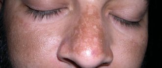
The main sign is a spot with uneven borders that does not hurt, does not itch, does not rise above the skin and does not peel off. There is no difference to the touch between healthy skin and a blemish. The color of the spot is all shades of brown, from light sand to brown, sometimes with a greenish tint. The sizes vary - from 1 cm to entire areas of the face.
Typical locations on the face are the forehead, bridge of the nose, area around the eyes, upper lip. The eyelids and chin remain intact. The spots, as a rule, are isolated and located one by one, but in some patients they merge, forming a continuous zone of pigment. The shape of the spots is uneven, sometimes bizarre, there is no symmetry.
The symptoms of chloasma do not threaten health in any way, but are a cosmetic defect. People with age spots on their faces often experience deep psychological discomfort.
Girls have special forms called perioral dermatosis. The spots are located symmetrically around the circumference of the mouth. These formations are particularly persistent and chronic. For many people, pigmentation spreads to the nasolabial folds over time.
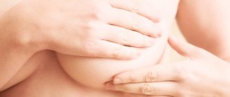
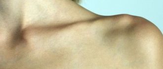
Sometimes uneven spots appear on the inner surface of the thighs, in the middle of the abdomen, and on the chest. Scars after wound healing, especially rough ones, can become pigmented.
Another type of pigment accumulation is dyschromia, or a line up to one cm wide, starting at the upper edge of the forehead and running obliquely across the cheek to the side of the neck. Such pigmentation often forms in severe diseases of the central nervous system: syringomyelia (cavities in the spinal cord), syphilis, Parkinson's disease.
People of Asian race develop bronze-colored spots.
During the period of active sun - spring and summer - the spots are more visible; in winter they fade somewhat, but never disappear completely. Once formed, the stain lasts for years, sometimes until the end of life. Chloasma may go away on its own after childbirth, but this does not always happen. With age, the number and color of age spots intensify.
At CELT you can consult a dermatologist.
- Initial consultation – 3,500
- Repeated consultation – 2,300
Make an appointment
Diagnostics
In many cases, diagnosis is not difficult. The typical location of the spots, their appearance and characteristics leave no doubt about the diagnosis. Chloasma is also supported by the fact that at birth there were no spots or inflammations on the skin.
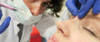
To confirm the diagnosis, two auxiliary methods are used: dermatoscopy and siascopy. Dermatoscopy is an examination using a special device under high magnification in good lighting. The procedure is completely safe and painless. First, gel or oil is applied to the stain so that light can penetrate through the stratum corneum. The turned on dermatoscope performs fluorescent illumination and multiple magnification, from 10 to 30 times, at which pictures are taken. Analysis of images provides answers to many questions.
Siascopy is a spectrophotometric instrumental study that allows you to find out the exact structure of pigment formation. The result is stored digitally for later comparison.
Clarifying the characteristics of a pigment spot is only part of the diagnosis. It is necessary to detect all changes that led to the formation of pigment. This is a complex task that requires careful examination.
It is recommended to perform the following steps and additional research:
- consultation with a gynecologist to assess hormonal status;
- Ultrasound of the pelvic organs and other organs according to indications;
- biochemical blood test with expanded liver complex;
- stool analysis for dysbacteriosis;
- coprogram (general stool analysis);
- Ultrasound of the abdominal organs;
- gastroscopy.
If diseases are detected during the examination, then consultation with specialized specialists and additional instrumental and laboratory diagnostic methods are necessary. But you need to start the examination with a visit to a dermatovenerologist.
Why does the skin turn yellow?
One of the most common liver syndromes, and quite striking, is a disorder of pigment metabolism - jaundice. The term “jaundice” refers to an increase in the level of bilirubin in the blood serum with its deposition in tissues and their icteric staining. Normally, its concentration is below 20 µmol/l (1.2 mg/dl). Jaundice staining of the skin and mucous membrane is visually diagnosed when the level of bilirubin in the blood is above 40 µmol/l.
Content:
- Why does the skin turn yellow?
- Causes and symptoms of yellowing skin
- When should you see a doctor?
- “Bad” and “good” yellowness of the skin
Artificial lighting can mask jaundice even at higher bilirubin levels. If the patient’s general condition is good, “false jaundice” should be immediately excluded. Excessive consumption of carrots, especially their juice, pumpkin, tangerines and some other plant products rich in xanthines, and the use of picric acid can cause icteric discoloration of the skin, especially the palms. The color of the sclera, mucous membrane and urine almost never changes. Biochemical examination showed bilirubin levels within normal limits.