There are contraindications. Specialist consultation is required.
Signs Causes of development Treatment methods Preparation for removal How is removal done? Indications Contraindications Recovery
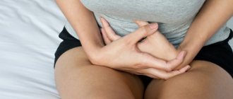
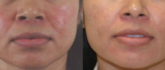
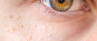
Spider veins , also called "telangiectasias", are the outward manifestation of dilated capillaries in the skin. They may have a bluish tint or dark red.
Capillaries can expand on any part of the body. Most often - on the face and legs. Appearance varies from tree-like to dotted. Linear reddish telangiectasias most often appear on the face (cheeks, nose) and are called “rosacea”. Women are more susceptible to the disease.
This disease is often a serious aesthetic problem. The presence of “stars” on the legs can be a symptom of the formation of varicose veins.
Sclerotherapy
Operation time - 10 minutes
Hospital recovery - not required
Cost of the operation: from 4,050 rubles.
Reasons for appearance
The nature of the disease can be varied. Some prerequisites for the formation of spider veins:
- constant work on your feet;
- venous insufficiency in chronic form;
- vein diseases;
- diseases of the endocrine system;
- smoking;
- taking hormonal medications, including contraceptives;
- drinking alcohol in large quantities.
If the disease is not associated with venous insufficiency or other ailments (this is determined before the removal procedure), then treatment requires additional examination by specialized specialists.
Volkov Anton Maksimovich
Phlebologist, cardiovascular surgeon
Doctor of the highest qualification category
“Spider veins are often one of the signs of chronic venous disease. The most common reasons are: long-term sedentary work (for example, accountant, cashier), sedentary standing work (for example, security guard, surgeon) and a combination of these reasons (for example, teacher, office employee). In this case, the patient will often feel heaviness in the legs in the evening, weakness, anxiety, pain in the legs and itchy skin. All of the above significantly reduces a person’s quality of life. Based on international recommendations, if you have at least one symptom, I advise you to immediately consult a specialist for diagnosis and treatment.”
Features of the vascular network on the chest
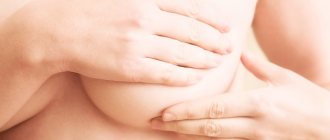
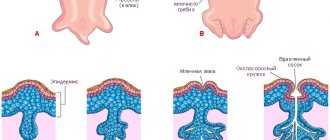
Most often, stars in the chest area occur in pregnant women. Similar modifications are associated with most conditions, and the formation of the fetus brings changes to the mammary glands. The expectant mother's breasts increase significantly in volume, and the blood supply system at this time begins intensive work in this area. The processes contribute to the appearance of a vascular network.
The mechanism of change occurs against the background of hormonal changes - after conception, a lot of estrogen and progesterone appear in the blood fluid, which directly support the progress of pregnancy. The restructuring of the body is accompanied by painful sensations in the chest area; they disappear after the first trimester of bearing a child.
It is important to choose underwear wisely
You can eliminate the feeling of chest discomfort during pregnancy by following these rules:
- Choosing the right underwear is an important nuance for such a crucial period. A comfortable bra ensures the healthy condition of the mammary glands;
- if pain intensifies or you feel lumps in the chest, you should consult a doctor for proper treatment of the disease;
- During pregnancy, you need to carefully select food products and avoid eating foods that provoke changes in the well-being of the expectant mother.
The use of cosmetic products for stretch marks is permissible only after consultation with the doctor.
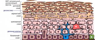
The skin surface becomes so dense that barriers to normal blood flow arise. An increase in pressure helps to normalize blood flow, which provokes stretching of the vascular walls and the formation of rosacea. Similar changes are also observed in liver pathologies: hepatitis, cirrhosis, liver cancer. Disruption of the pancreas and thyroid glands can also be accompanied by skin changes that appear on the walls of blood vessels. In such cases, the disease affects not only women, but also men.
Treatment and removal methods
Important! Spider veins cannot be removed with external ointments and gels. To solve the problem, you need to contact a surgeon who will provide effective medical care.
At SM-Clinic, patients are offered to remove spider veins on the face and other parts of the body using sclerotherapy.
The technique is aimed at “gluing” the walls of the capillaries and preventing blood from entering them. As a result, the vascular mesh will not be visible through the skin.
Removal of spider veins and spider veins is a painless and short procedure with quick healing and no scars.
Measures to prevent the disease
Venous mesh is formed due to various reasons. If there are no significant disorders in the body, this phenomenon can be eliminated by using cosmetic procedures and taking vitamin complexes. To protect the body from the appearance of unpleasant stars, you must adhere to the following principles:
- complete cessation of bad habits;
- maintaining a balanced diet;
- exercise;
- regular vitamin therapy;
- timely visit to the doctor.
The first thing you need to do is completely give up bad habits.
Taking care of the capillaries is of great importance. Modern drugs allow you to protect them from harmful influences, restore elasticity and firmness. An exceptionally comprehensive approach will ensure care for the body and the disappearance of unpleasant manifestations forever.
At times, the vascular network that forms does not pose a threat to the body. Their appearance may confirm the presence of venous pathologies. In such situations, it is necessary to take care of the condition of the blood vessels, strengthening them. The formation of large sections where veins are clearly visible becomes a significant reason to consult a doctor.
Description of the procedures
Today, there are several modern methods of sclerotherapy, which allow us to solve the issue of radical treatment of initial forms of varicose veins in a non-surgical way.
Compression sclerotherapy
It is a classic technique for the treatment of varicose saphenous veins. Allows you to remove the external manifestations of varicose veins, without affecting the cause of the disease - pathological reflux. Typically, compression sclerotherapy is prescribed after combined phlebectomy to eliminate remaining varicose veins or as an alternative to miniphlebectomy after correction of valvular insufficiency using non-surgical methods.
Foam-foam sclerotherapy
Microfoam sclerotherapy is a type of sclerotherapy that uses a drug in the form of very fine foam. The foam form allows you to create a volume of sclerosant several times greater than the volume of the original solution with the same content of the active substance, ensuring longer and more dense contact of the drug with the vein wall. This increases the reliability and effectiveness of the procedure while reducing the risk of complications.
ECHO sclerotherapy
This technique involves performing the sclerotherapy procedure under full ultrasound control. It is usually used for sclerosis of altered veins deep in the subcutaneous fat, incompetent perforating veins, and main trunks of the saphenous veins using a catheter technique.
Microsclerotherapy
Used to treat spider veins and reticular veins of the lower extremities. The essence of the procedure is multiple microinjections of sclerosant directly into problem areas. The effectiveness of the technique is quite high - in most cases, complete disappearance of the venous network is observed.
Catheter scleroobliteration
Designed for sclerosis of the trunk of the great and/or small saphenous vein. Those. This technique allows you to eliminate the main pathogenetic mechanism of varicose veins - ostial and stem reflux. A catheter is inserted into the lumen of the vein, through which sclerosant is supplied. Simultaneously with the supply of the drug, the catheter is moved along the vein and an elastic bandage is applied to the sclerotic areas. The technique is an alternative to surgery for radical removal of the subcutaneous line. Disadvantages include the possibility of use only in the early stages of varicose veins and lack of effectiveness.
Diagnosis of the disease
Determining treatment options for a venous network involves conducting an examination to identify the stage, level and volume of vascular pathology. Doppler ultrasound (USDG) is used as the main examination method, but other methods can be used according to indications. In addition to the phlebologist, the examination is carried out by a vascular surgeon, endocrinologist, gynecologist and cardiologist. If necessary, it is possible to prescribe auxiliary examinations to identify pathologies in the functioning of certain organs.
Rehabilitation after procedures
There is no need to stay in the hospital; patients can immediately return to their normal lifestyle. Restrictions include avoiding exposure to the sun for 2 weeks, visiting a bathhouse, and not massaging the operated area.
Contact SM-Clinic for quality treatment
In our clinic in St. Petersburg you can remove spider veins on the body and face using sclerotherapy (prices vary, they are listed on the website). We employ some of the best specialists in the Northern capital, and comfortable conditions have been created for patients. The clinic has advanced diagnostic and treatment equipment.
You can ask questions about prices, features of laser and other removal of spider veins on the legs and other areas by phone.
Make an appointment with a phlebologist - give yourself beauty and health.
Vascular network on the chest, nuances of occurrence
The venous web does not pose a danger to the body. Such changes can disappear on their own without causing discomfort to the person. However, there are situations of rapid growth of the mesh, which indicates the presence of disorders in the body. Then discovering the true etiology is quite difficult.
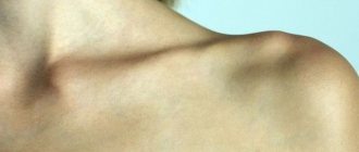
Rapid weight loss can cause previously hidden veins to bulge
Additional conditions that cause the development of a venous web in the thoracic region are:
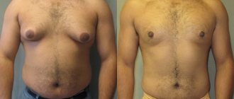
- Powerful physical stress on the body. The appearance of mesh in men can be triggered by taking drugs to stimulate muscle growth.
- Rapid weight loss. A sharp reduction in body weight can cause previously hidden veins to bulge. After gaining normal weight, the formations disappear on their own.
- Surgical intervention. Plastic correction of organs can cause the growth of the venous network.
- Excess weight provokes vascular disorders, as a result of which vascular cobwebs form on the body.
- Pathology of the liver and adrenal glands. The appearance of a mesh under the breast indicates significant disturbances in these organs.
Similar manifestations also occur with high blood pressure - unable to withstand the load, capillaries rupture and hematomas form. If the reason for the changes is known, there is no need to panic. Otherwise, the help of a doctor is required, who will inform you about all the intricacies of the pathology and suggest the correct method of therapy.
Prevention of an abnormal phenomenon
If the venous network on the chest is caused by non-pathological factors, vitamin complexes containing components that can restore and increase the tone of the vascular walls will help get rid of it.
To avoid re-appearance of stars, experts recommend:
- Avoid chest trauma.
- Quit smoking and alcohol.
- Do not get carried away with spices and strong coffee.
- Make therapeutic masks for the skin of the chest and décolleté (not heating), regularly use nourishing lotions or cream.
- Lead an active and healthy life, avoiding increased physical activity.
- Avoid hypothermia.
- Get rid of excess weight.
- Stick to proper nutrition.
- Monitor blood pressure levels.
- Avoid stressful situations.
- Make time for rest.
- Eat nutritiously and variedly.
- Wear comfortable underwear.
- Take a contrast shower.
- Stick to your daily water intake.
- When working sedentarily, do special exercises aimed at preventing stagnation of blood in the vessels.
Diagnostics
To determine an accurate diagnosis, to find out the type and degree of pathology, a complex of studies is required, which is carried out by both a mammologist and a phlebologist (it is possible to involve a therapist or oncologist). First, doctors collect information (by questioning) about how the disease progresses, what symptoms besides protruding veins bother the woman, whether there are hereditary preconditions, or risk factors for developing the disease.
After studying the medical history, instrumental diagnostics are performed. A frequently used method is color duplex examination. It allows you to measure the speed of venous blood flow, map blood vessels and identify the exact location of points where their lumen is narrowed due to blood clots. X-ray examination (phlebography) shows the shape of the veins, as well as the functioning of the valves and the direction of blood flow.
Since often visible veins indicate neoplasms in the breast, the mammologist must prescribe a test for inflammation and tumors. To do this, a blood test is taken for tumor markers, ultrasound, radiography, and ductography (mammography with a contrast agent) are done. If lumps are found in or near the mammary glands, a biopsy is performed followed by examination of the cellular material.