Indications for lipofilling of the legs
- Disproportional distribution of soft tissues;
- Defects of soft tissues of the legs;
- Post-traumatic deformity of the legs;
- Asymmetry of the legs;
- Insufficient volume of the inner part of the lower leg;
- Visual curvature of the legs without bone deformation.
The lipofilling technique involves taking adipose tissue from areas of excess and transferring it to places where volume needs to be added.
Removing Polyacrylamide Gel
The past years have revealed the danger of injecting polyacrylamide gel into soft tissues. The gel migrates, causes visible deformations, provokes the development of inflammation and trophic disorders. All this requires the removal of the previously introduced gel ( more " ).
Indications for polyacrylamide gel removal
Presence of polyacrylamide gel in soft tissues
Our recommendations
For more detailed information about how polyacrylamide gel removal using the described method can help you, about the indications and contraindications of the method, we recommend that you consult a plastic surgeon with experience in PAAG removal.
How is lipofilling surgery performed?
At the initial consultation, the surgeon conducts an examination and makes sure that the visual curvature of the patient’s legs is not associated with skeletal deformation. It is also important that the legs are not affected by varicose veins, since this disease is a contraindication for lipofilling of the legs.
Then the doctor calculates the amount of fat tissue required for transplantation and discusses with the patient from which areas the donor fat cells will be taken. The undoubted advantage of the technique is that, as a bonus, you can correct problem areas - the sides or back, taking the required amount of fat from them.
The operation is performed under both local and general anesthesia. The procedure is divided into several stages:
- sampling of adipose tissue using special cannulas;
- processing of cells before injection into the legs;
- uniform filling of problem areas.
After the operation, the patient is placed in a comfortable hospital room. If the operation was performed under local anesthesia, then within a few hours after the surgeon’s examination he is allowed to go home.
The rehabilitation period lasts about ten days. During this period, the swelling completely goes away and the puncture sites heal. The surgeon evaluates the final result after several months, when the fat cells have completely taken root.
Contraindications
This correction technique has standard contraindications associated with surgical intervention and putting the patient under general anesthesia. Permanent and temporary (go away on their own or can be eliminated with treatment) contraindications:
- diabetes;
- oncological processes;
- blood clotting disorder;
- severe pathological processes in the body;
- acute bacterial infections and viruses;
- period of menstruation in women.
Restrictions during the rehabilitation period
You must wear compression tights for a month after the procedure. It is also necessary to exclude:
- long walks for a month;
- active sports for a month;
- all types of hair removal, including shaving, for 2 weeks;
- visiting the beach, sauna and solarium for a month;
- massage of the legs for 3–6 months.
Structural anomalies
From an orthopedic point of view, straight human legs are defined as the correct relationship of four points (mid-thigh, lateral calf, calcaneus, and knee meniscus) in a standing position that forms a straight line without any deviation.
In case of false and true curvatures, special plastic surgery of the lower leg will help hide the defect; in Moscow, cruroplasty is performed by the plastic surgeon Gulyaev.
The first anomalies in the structure of the legs are formed in childhood, with the beginning of walking. An X-shaped or arched curvature of the legs develops, which is accompanied not only by an unattractive appearance, but also by pain and discomfort when walking.
An arcuate curvature is an outward deviation of the knees; the distance between the menisci is much greater than normal, forming an oval. A woman’s appearance immediately loses its aesthetics; training and exercise are unable to hide severe joint and bone pathology.
X-shaped curvature of the limbs is accompanied by a decrease in the distance between the menisci. This pathology is a characteristic symptom of rickets in childhood. As the child grows, in the complete absence of treatment, the altered bones and joints undergo sclerotic changes, and this aggravates the disease and irreversibly deforms the limbs.
False anomalies occur with atrophic changes in muscle tissue, low body mass index, as well as genetically determined too weak differentiation of myocytes. A thin layer of muscles creates the effect of thin legs; due to the rounded joints of the knees, this can easily give the impression of a so-called false curvature.
Advantages of the lipofilling method
If we compare the lipofilling method with leg shape correction using implants, it certainly has a number of advantages:
- lipofilling is carried out without skin incisions, the method is low-traumatic, the pain syndrome is less pronounced;
- after lipofilling there are no scars left;
- the use of the patient’s own adipose tissue eliminates the risk of non-survival of adipose tissue implants;
- it takes root easily, the lower leg remains soft, since the adipose tissue has a homogeneous structure;
- recovery takes place in a short time and with minimal restrictions;
- the result of lipofilling is long-lasting and durable;
- bonus: adipose tissue for lipofilling is taken from those areas where it is in excess - two zones are corrected in one procedure.
Cruroplasty: preparation
Before scheduling an operation, a woman must undergo a comprehensive examination with various laboratory tests, instrumental studies and consultations with doctors. The latter allows you to diagnose exacerbations of any concomitant pathology, which can negatively affect recovery after cruroplasty.
In the presence of congenital anomalies in the structure of bones and joints, consultation with a neurologist and orthopedist is indicated. The doctor examines the calf muscles, ankle and knee joints, determines the level of sclerotic changes, and also excludes the presence of exacerbations of arthrosis and arthritis.
Laboratory tests are needed to identify chronic or acute inflammation, pathology of hemostasis with increased bleeding, and various disorders of thrombus formation. Instrumental examination methods include ultrasound examination of muscle fibers if the patient has suffered myodystrophic diseases or injuries.
Doppler examination of blood vessels makes it possible to detect chronic venous pathology, thrombophlebitis, varicose veins, and atherosclerotic changes in the patient. Doppler is performed a week before surgery and a week after it in order to eliminate the risk of thrombus formation in large venous trunks, as well as pathology of the valve apparatus.
Before the operation itself, the patient limits the consumption of alcoholic beverages and smoking, because all this has a bad effect on recovery, worsening the overall prognosis. Doctors advise excluding starchy and sweet foods from food a couple of weeks before surgery in order to reduce the likelihood of bacterial and mycotic flora joining.
After a full examination, in the absence of contraindications, the patient undergoes cruroplasty, the price of which is determined by the techniques and implants used.
During the consultation before the operation, the doctor, if necessary, creates a three-dimensional image of the desired result, taking into account the wishes of the patient, using a computer. The height of the bend, the severity of the side ridges and contour are determined.
The result of shin contouring is a harmonious and aesthetic appearance of the legs.
Benefits of lipofilling of the legs at the UMG center
Specialization
Lipofilling is one of the important areas of the UMG Center for Aesthetic Medicine. Over the past year, the center has performed more than 750 successful lipofilling procedures.
Skill level
The head of the surgical department of the UMG Center for Aesthetic Medicine, Evgeniy Igorevich Savelyev, defended his PhD thesis on the topic “Lipofilling of the legs” and is one of the leading Russian experts in this field.
Equipment
The equipment of the UMG center corresponds to the level of the best clinics in Europe and America. To collect fat cells, the center uses an expensive Vazer device, as well as a Body-Jet water-jet liposuction system. Such equipment is used today in only ten Russian clinics, and the results obtained with its help evoke rave reviews from patients and doctors.
Endoprosthetics: technique
Leg surgery is performed under general endotracheal anesthesia with epidural anesthesia for the patient. This allows you to achieve complete relaxation of the deep muscle fibers and prevent displacement of the implant in the postoperative period. Before the operation, the doctor marks the area where the implant will be installed with a marker to ensure complete symmetry.
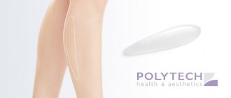
Endoprosthetics is a unique operation that instantly restores the shape and contour of the entire calf area. Plastic surgery of the lower leg using endoprostheses is performed using several approaches:
- Submuscular
- Subfascial.
Subfascial method
An incision is made in the physiological skin fold under the knee; with the help of blunt bougienage, an artificial pocket is created under the connective tissue membrane of the calf muscle bundles. A small silicone implant is inserted into the pocket and secured with stop ligatures in the upper part of the popliteal aponeurosis.
Submuscular method
This method is designed specifically for the use of full-shell semilunar implants, which require very strong fixation. Soft tissues, tendons and ligaments tightly surround the endoprosthesis, thus preventing its movement. The incision is made in the popliteal region of a larger size than with subfascial endoprosthetics.
A pocket is formed under the deep fibers of the calf muscles, and an endoprosthesis is installed there. The tension of the muscle plates leads to reliable retention of the endoprosthesis and eliminates the possibility of displacement.
If the tension of the muscle fibers is weak, cruropexy is performed using resection and suturing of connective tissue bundles, which are attached to the places of anatomical attachment, as well as to the aponeurotic popliteal cord.
The submuscular method is more often used due to the creation of the desired shape without the risk of excessive contouring of the implant. Today's endoprostheses for cruroplasty interact with surrounding tissues, succumbing to the resistance of muscle fibers, and take on a physiological shape at the time of active movements. The results of cruroplasty after submuscular fixation are always distinguished by their naturalness, as well as the rapid transformation of the contour of the legs and the shape of the calf muscles.
When the doctor installs implants on both legs, he will definitely compare the position and strength of the prosthesis. After this, fascia, tendons, aponeuroses, and muscle fibers are sutured layer by layer. The epidermis is sutured with a transcutaneous cosmetic suture, which heals with minimal defects.
