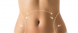
Hyaluronic acid fillers are most often used for contouring due to their high safety profile. Despite this, no practitioner is immune from serious complications of injection correction. The article by estet-portal.com presents a case of tissue necrosis of the upper lip after injection of HA filler from the practice of doctors Peter Hirsch, Manfred Infanger and Armin Kraus. The authors talk about the causes of necrosis after injection of fillers , and also provide recommendations for the management and prevention of this complication .
- Case study: tissue necrosis of the upper lip after HA injections
- Tissue necrosis after injection of fillers: causes, therapy and prevention of complications
Some statistics
Today, the percentage of undesirable consequences from appearance correction using fillers is negligible. According to various sources, it ranges from 1 to 5%. But just a couple of decades ago, this figure was several times higher - then the quality of “beauty injections” was not so perfect, and the training of doctors was not so perfect. Currently, most fillers are biocompatible, that is, they do not cause allergies, and specialists working with them are required to have a certificate indicating that they have completed training in the use of this substance.
What complications arise after contouring?
- Strong pain.
It is caused by the introduction of any foreign substance under the skin. How to get rid of it? In this case, discomfort is easier to prevent. To do this, ask your doctor to select a filler with lidocaine. It reduces discomfort and also reduces swelling. - Swelling and hematomas.
This is a common reaction to contouring. For some it is more pronounced, for others less. Usually the skin condition returns to normal within 24 to 36 hours.How to minimize? Let's start again with prevention. We recommend taking medications that strengthen blood vessels in advance. If complications after contouring have already appeared, special ointments should be applied to the injured area. Your doctor will select the best cosmetic product for you.
- Granulomas and fibrous capsules.
Inflammation of tissues is their protective reaction to an external irritant, especially if hygiene rules are not followed. The filler can also “wake up” infections dormant in the body. As a result, nodular seals may appear at the injection sites. Small ones are called granulomas, and large ones are called fibrous capsules.What to do? Granuloma can be eliminated with the help of advances in hardware and injection cosmetology. In particular, laser therapy and plasmafilling, peeling or dermabrasion. To prevent small compactions from turning into fibrous capsules, you will have to take anti-inflammatory drugs. And if the inflammation has already grown, you will have to consult a surgeon.
- Allergic reaction.
It is caused by individual intolerance to the drug used. What to do? Urgently inject a corticosteroid drug. - Gel migration or protrusion.
The drug changes location, distorting the proportions of the face if it was injected too deeply. If, on the contrary, the filler is placed too superficially, unevenness and bumps will form on the treated area. How to get rid of it? In both cases, another session of microinjections is required. The doctor must administer a drug whose components will break down the filler. However, this measure is effective only when using “beauty injections” based on hyaluronic acid. If you were given something else, correction of complications will take an indefinite amount of time. - Compression of blood vessels.
This leads to swelling and unhealthy redness of the tissue, and subsequently to necrosis with scarring. The reason for such consequences is the use of a low-viscosity filler, which accidentally enters the lumen of the vessel, or the doctor’s injection of a high-density drug too deeply. What to do? If blood vessels are compressed, the doctor prescribes symptomatic therapy.
One of the complications of contour plastic surgery is compression-ischemic syndrome, the consequences of which can be irreversible. Why does this complication occur? How to avoid it? And what should a doctor do if it does begin to develop?
In the list of complications caused by the introduction of both permanent and biodegradable fillers, compression-ischemic syndrome (CIS) is not the last place. Of course, a lot depends on the physicochemical and volumetric properties of the drug used, as well as on its quality. However, an equal role, if not the main one, is played by the professionalism of the doctor. In most cases, it is a medical error resulting from neglect or ignorance of the features of the layered anatomy of the face or a violation of the filler injection technique that can explain the compression-ischemic syndrome. It is caused by two main reasons:
1) injection of the drug directly into a blood vessel and, as a result, thrombosis;
2) injecting an excessive amount of the drug close to the vessel, which can cause compression.
As a result, the trophism of soft tissues is disrupted, and ischemia (necrosis) occurs as the most severe form of this complication.
Depending on how quickly the patient seeks help and the doctor begins to carry out rehabilitation measures, the effectiveness of the therapy depends. It should be remembered that the clinical picture of compression-ischemic syndrome manifests itself soon after the contouring procedure with an increase in edema, a change in skin color at the site of filler injection, and severe pain.
There are two types of CIS: arterial and venous. In the first case, the skin becomes pale, in the second, the venous pattern intensifies. Let us recall that necrosis (death of cells and tissues in a living organism) has four stages of development:
1. Paranecrosis (from the Greek para - near and nekros - dead) is a set of reversible nonspecific changes in living cells that occur in response to the action of various damaging agents.
2. Necrobiosis (from the Greek nekros - dead and bios - life) - changes in a cell or tissue that precede its death.
3. Apoptosis (from the Greek apo - without, from, from and ptosis - fall, death, dying, “falling of leaves”) - the process of programmed cell death; a regulated process of self-destruction at the cellular level.
4. Autolysis (from the Greek autos - itself and lysis - dissolution, destruction) - the breakdown of cells and tissues of the body under the influence of the hydrolytic enzymes they contain.
Stages of necrosis: 1 – paranecrosis, 2 – necrobiosis, 3 – apoptosis, 4 – autolysis.
When the process of tissue necrosis reaches apoptosis, it is considered irreversible and the appearance of aesthetic defects cannot be avoided. Let's look at several examples of compression-ischemic syndrome in those areas of the face where contouring is most often used.
Buccal region
Vessels aa are located in this zone. facialis (facial artery), transversa faciei (transverse artery of the face), a. infraorbitalis (infraorbital artery). The facial artery lies relatively superficially in the projection of the anteroinferior edge of the masticatory muscle (masseter), at the point of inflection over the edge of the lower jaw, and near the alar cartilage of the nose - in the thickness of the pancreas. And in other areas of the face aa. facialis is located in the spaces between the facial muscles.
Photo 1. Patient K., at the time of treatment, soft tissue necrosis at the apoptosis stage was diagnosed, caused by compression of aa. facialis and a.,v.,n. infraorbitalis.
Patient K. (photo 1) came to the clinic with an increasing clinical picture of CIS after contour injection plastic surgery with a drug based on hyaluronic acid. The clinical picture is due to compression of the vessels located in this area by the gel, which occurred due to excessively deep injection of the filler. At the time of treatment, soft tissue necrosis at the apoptosis stage was diagnosed. The patient was urgently administered hyaluronidase and prescribed long-term vasoactive and antibacterial therapy. Today she is undergoing a course of fractional laser therapy to correct residual scarring of the skin.
Photo 2. Patient V., after repeated administration of calcium hydroxyapatite, required surgical intervention to remove it.
Patient V. (photo 2) successfully underwent contour plastic surgery of the nasobuccal sulcus using calcium hydroxyapatite. The complication arose after the additional correction procedure. Fortunately, the patient sought help just two days after the filler injection, when she felt pain in the cheek area, saw increasing swelling and increasing redness, and a local increase in temperature. A computer study showed an accumulation of gel in the area of the exit of the neurovascular bundle (a., v., n. infraorbitalis), causing compression. Unlike fillers based on hyaluronic acid, preparations with calcium hydroxyapatite do not have an antidote, so the only way to remove the gel and relieve compression from a., v., n. infraorbitalis – surgical. Antibacterial and vasoactive therapy was then prescribed.
Photo 3. Patients after injection of HA-based filler with a complication in the form of compression-ischemic syndrome caused by compression of the angular artery. Seeking help a few hours after the procedure allowed us to avoid full-thickness tissue necrosis.
Nasolabial area
In this area, the danger is compression of the angular artery (photo 3), which is the terminal branch of the facial artery, runs up the lateral surface of the nose, giving small branches to the back and wing of the nose.
Zygomatic region
This area is quite safe for a doctor to work from the point of view of the likelihood of introducing the gel directly into the vessel and the formation of thrombosis, since large vessels do not pass through here. All complications arising in this area are caused by overcorrection, that is, an excessive amount of the administered drug, and, as a consequence, compression of the blood vessels.
Periorbital region
Recently, contour plastic surgery in the periorbital zone has become very popular.
Here, the most dangerous area is considered to be the area of the inner corner of the eye, where the angular artery anastomoses with the dorsal nasal artery, a branch of the ophthalmic artery. Cases of vision loss due to improper filler injection have been described in the literature.
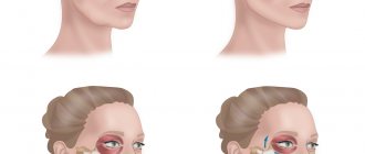
Photo 4a - Fibrous tissue under the lower eyelid resulting from the injection of an excessive amount of permanent gel. Photo 4b - Compression-ischemic syndrome of the supraorbital artery.
Patient M. (Figure 4a) had an excessive amount of permanent gel injected between the skin and muscle, which caused the formation of fibrous tissue and scars. To reduce their severity and restore the skin, fractional laser therapy was prescribed.
Patient S. (photo 4b) requested additional correction of vertical forehead wrinkles with a hyaluronic acid preparation.
15 years ago, she had a permanent filler injected into this area, which provoked the formation of fibrous tissue. Injections of HA filler caused compression-ischemic syndrome of the supraorbital artery precisely because of the presence of this tissue in the forehead area. It should be understood that the administration of the HA drug to patients who were once injected with permanent filler is undesirable, since there are risks of complications.
This is especially true for fillers based on polyacrylamide gel. In all such cases, it is safer to carry out correction using lipofilling or plasmafilling.
Frontal region
In this area, the supratrochlear and supraorbital arteries pose a danger. The supratrochlear artery is one of the terminal branches of the ophthalmic artery and is located medially to the supraorbital artery. It goes around the supraorbital margin and goes upward, supplying the skin of the medial parts of the forehead and muscles.
Photo 5. Patient R., resolution of clinical symptoms of layered tissue necrosis under the influence of combination therapy: a – before therapy (2 weeks after filler injection); b – 2 weeks after the start of treatment for CIS; c – 4 weeks after the start of treatment for CIS; d – final result of CIS therapy.
Patient R. (photo 5) came to the clinic with full-thickness tissue necrosis in the forehead area, which developed within a few days after the use of HA filler. She was administered hyaluronidase and prescribed a course of antibacterial and vasoactive therapy to improve microcirculation.
A month and a half after the start of treatment, all the crusts disappeared, the pustular phenomena resolved, complete tissue regeneration occurred, but the depigmented scar still remained.
Lip area
In this area, the lower and upper labial arteries pose a danger - they are quite large. A needle is most often used to inject filler here, so the complication is usually associated with thrombosis, that is, the drug entering directly into the vessel.
Photo 6. Thrombosis of the superior labial artery: a – a few days after the injection of HA filler; b – one month after the start of treatment for CIS; c – one year after filler injection.
Patient M. (photo 6) came to the clinic a few days after the procedure for introducing HA filler. The complication was caused by the gel entering directly into the vessel. Timely treatment (administration of hyaluronidase, vasoactive and antibacterial therapy) made it possible to cope with the situation with virtually no consequences, that is, without visible changes in the skin.
Photo 7. a – development of compression-ischemic syndrome of the labial artery upward, to the area of the facial artery; b – thrombosis of the labial artery (MRI image).
Patient L. (photo 7) with compression-ischemic syndrome of the upper lip caused by thrombosis of the superior labial artery, came to the clinic a week after the procedure - during this time she did not receive any treatment. Further spread of ischemia moved to the area of the facial artery. Emergency treatment included the administration of hyaluronidase, anticoagulant and vasoactive therapy, non-steroidal anti-inflammatory drugs, and antibacterial drugs. Homeopathic medicines were used locally to improve microcirculation.
After a month of treatment, it was possible to achieve resolution of the clinical picture in the nose and nasolabial area, however, the necrotic process in the upper lip area still persists, which will require surgical intervention to restore the upper lip. It is important for the doctor to understand that if compression-ischemic syndrome is suspected, it is necessary to act as quickly as possible.
Emergency help for complications after the injection of HA-based filler is the introduction of hyaluronidase and the use of nitroglycerin in a spray to relieve vasospasm. Then, focusing on the severity of the situation, the patient must be prescribed anticoagulants, vasoactive therapy, nonsteroidal anti-inflammatory drugs, dextrans, antibacterial therapy, and homeopathic medications. After this, therapy using fractional or CO2 laser.
If a complication occurs after the injection of another filler, the patient should be referred to a surgeon.
Olga Danishchuk, plastic surgeon, assistant at the Department of Surgery, IPK FMBA of Russia; Elena Karpova, MD, plastic surgeon, member of the American Academy of Facial Plastic and Reconstructive Surgery (AAFPRS), member of the Society of Plastic, Reconstructive and Aesthetic Surgeons (OPRESH) of Russia, Moscow
How to look beautiful and young without unpleasant consequences?
Once again, we draw your attention to the fact that the percentage of complications after “beauty injections” is very small. To minimize the risk, you should adhere to the following rules:
- Choose a well-known clinic. Don’t play roulette by agreeing to lucrative offers from people who practice contouring at home.
- Don't hide information about your health. The doctor needs to know if you have allergies, a tendency to edema, etc.
- After the specialist selects the filler, read the information about this drug - it must be certified in Russia. If necessary, ask to replace the proposed gel with a safer one.
- Make sure that the dermatocosmetologist has a certificate confirming the right to work with this substance.
The VERSUA Clinic team wishes you to remain young and beautiful for many years to come!
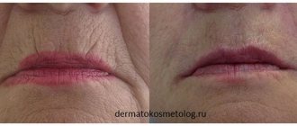
Case study: tissue necrosis of the upper lip after HA injections
The patient, 19 years old, came to the authors' clinic one week after having a filler injection into her upper lip. It is unknown which product was administered to the patient and in what quantities. According to the patient, the procedure was carried out after applying an anesthetic cream. Despite this, during the injection the girl felt a strong burning pain that radiated from the left side of her upper lip to her nose. Apart from cooling compresses, no specific therapy was prescribed after contouring.
Follow us on Instagram!
During the examination, a dry necrotic area measuring 8 x 10 mm was discovered, as well as swelling of the left side of the upper lip . A blue line was visualized from the upper lip through the philtrum and columella to the tip of the nose.
Photo 1: partial necrosis of the left upper lip and impaired skin perfusion of the philtrum, columella and nasal tip after HA injection
Therapy
Since a long time had passed since the procedure, the administration of hyaluronidase was inappropriate. The authors decided to remove the maximum possible amount of filler located under the area of tissue necrosis.
The patient underwent an infraorbital nerve block, a small stab incision was made on the side of the mucosa, and the filler was removed.
You may also be interested in: A case of occlusion of the posterior ciliary artery after the injection of hyaluronic acid into the forehead area
As topical therapy, dexpanthenol ointment was prescribed, as well as regular application of dry dressings for 2 weeks.
Photo 3: filler removal after infraorbital nerve block
After 3 months , the wound healed, and a small scar formed in its place. There was also significant volume loss in the left upper lip. The skin color of the affected areas was almost restored to normal, but a thickening was felt in the tissues, which indicated subdermal scarring. The asymmetry was enhanced by the presence of filler on the right side of the upper lip.