One of the new ways of introducing fillers has become Jolie angles
.
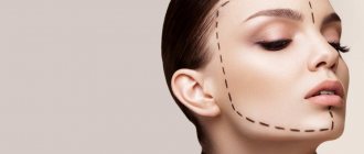
Angelina Jolie has been the ideal of many women for many years. Millions of ladies are trying to imitate her and want to look like Jolie. Many people ask the cosmetologist for lips like Jolie, a nose like Jolie, and very often ladies want a lower jaw like Jolie. Unfortunately, many women are deprived of a clear contour of the lower jaw.
For most women, these angles are completely absent from birth. Also, with age, the contour of the face floats, and the contours of the lower jaw lose their clarity and expressiveness, and sag greatly. Such age-related changes must be corrected. A clearly defined lower jaw adds expressiveness to the entire face
, the facial contour becomes clearer. Modern cosmetology allows you to achieve an ideal facial contour.
This procedure has another name, which is used more rarely - angles of youth. It is called so because these angles are characteristic of young girls; such angles give the face youth and femininity, as well as sophistication.
If we turn to the aesthetic side of the issue, then with the help of such angles you create ideal facial proportions. There is a triangle of youth, which includes a well-defined chin. You can see these proportions in Angelina Jolie.
The procedure is great for those who have a thin face. Unfortunately, if you have chubby cheeks, this procedure is undesirable - the face may take on a rougher and more massive appearance.
Duration of the procedure and result
This technique of introducing fillers has gained popularity in the United States. This is where Angelina Jolie is a role model. Recently, the technique has begun to gain popularity in our country. The drug is injected into the jawline and into the very corners of the lower jaw
. No one will notice that you have been injected with drugs. The result will look like it is your own bone.
Naturally, this method of introducing fillers began to be used after a large number of tests. Cosmetologists have been studying this technique for a long time, achieving the most ideal result. The procedure takes very little time and allows you to achieve the most impressive results in the shortest time.
What problems can be solved
The main tasks that the procedure allows to cope with are replenishing lost volume, changing the size and shape of the angles of the jaw, restoring the asymmetrical position, eliminating pronounced creases, folds and wrinkles in the lower third of the face. Effects that can be achieved:
- the jaw becomes clearly defined;
- the face acquires a correct and proportional shape;
- a protruding chin becomes less noticeable;
- if the chin is too sunken, then it acquires the correct size and increases;
- the double chin disappears if the person is of normal weight;
- your appearance will be harmonious and attractive.
Reviews
Advantages of the Jolie angles procedure
This procedure has many advantages.
- Firstly, you do not have to contact surgeons and undergo surgical intervention. Everything is done in a non-surgical manner. This procedure is most often done with hyaluronic acid. This drug takes root well in the body, and the possibility of drug rejection is excluded. Hyaluronic acid is found naturally in the body.
- Secondly, you don't have to go through rehabilitation. The result is visible almost immediately; there is no need to maintain it. It is enough to carry out the procedure once every couple of years. The effect of this procedure lasts about 2 years. After the procedure, you will not receive a single scar or traces of intervention. The fillers already contain an anesthetic, so the procedure is painless. If necessary, the filler injection area is additionally anesthetized, so you will not experience any unpleasant sensations.
With just one filler injection session, you will get a completely new jaw line and defined angles.
The procedure will help those who have asymmetry
, and who simply want to make their face more defined.
| When you sign up online, you not only receive a discount on the service, but also an individual consultation with a leading specialist for free! | Sign up online 5% discount |
What to do after the procedure
You can immediately return to your normal lifestyle - you will look the same as always, only better, and you won’t have to sit at home. There are some restrictions - in the first few days it is not recommended to sunbathe or visit the sauna or bathhouse. Chin contouring in men has its own characteristics. During the healing period of the piercings, it is recommended, if possible, to refrain from shaving for a couple of days.
In the first couple of days, there may be some swelling at the injection sites, and sometimes (rarely) small bruises. They go away on their own within a few days.
Contraindications
The procedure has practically no contraindications, but it is undesirable to carry out the procedure during pregnancy, cancer, inflammatory processes, and allergic reactions. You can learn more about contraindications in consultation with a cosmetologist, which must be carried out before the procedure.
No special preparation is needed for the procedure, but drinking alcohol before the procedure is not recommended. A cosmetologist will solve all your questions. He will tell you whether you can have the procedure and how to prepare for it.
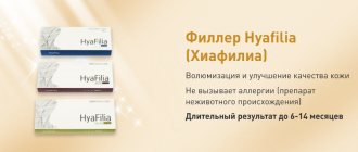
A variety of fillers are used to create Jolie's angles. They are made based on hyaluronic acid.
The main drugs used for jaw contouring
- RESTYLANE is an effective and comfortable gel, does not cause inflammatory reactions, its use is extremely rarely accompanied by swelling, and gives a clear, graphic contour of the lower jaw.
- TEOSYAL is a filler with minimal protein content, safe, hypoallergenic.
- JUVEDERM ULTRA - preparations with hyaluronic acid, which contain lidocaine, are distributed as evenly as possible under the skin. The wide range allows for both light correction and pronounced volumetric correction of the jaw and chin.
Carrying out the Jolie corners procedure
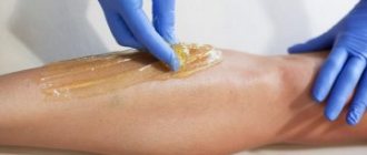
The face must be cleansed of cosmetics, fat, and dust. The skin is treated with antiseptic agents to prevent bacteria. The selected area is anesthetized to ensure that the procedure is as painless as possible.
Additional anesthesia is applied at your request. It all depends on how much pain you feel. The area of drug administration is marked with a special marker so that it is clear where the drug should be injected. Then the drug is injected, the skin is disinfected, and the contour is modeled.
After the procedure, you cannot knead the filler yourself with your hands, as you can seriously ruin the result. It is advisable not to visit baths and saunas, and also not to physically influence the area where fillers are introduced.
Conduct a free consultation
and the procedure itself for correcting the angles of the lower jaw can be done at the cosmetology and dentistry clinic of Dr. Korchagina.
Read more about the procedure and medications here...
Promotion!
Correction of the angles of the lower jaw “Jolie Profile”
Cost: from 15,000 rub.
instead of 18,500 rub. Order a service Ask a question
Main advantages:
- Minimum tissue damage;
- High efficiency:
- No bruising or swelling, no need for long-term rehabilitation;
- Affordable cost: compared to plastic surgery, this intervention is much cheaper.
Perhaps the only disadvantage of correcting jaw angles with fillers is the temporary effect. The gel, which is administered by injection, gradually dissolves and is excreted from the body. The pronounced effect lasts from a year to two; if necessary, the injections can be repeated - it is safe.
Augmentation of the jaw ridge using block allogeneic bone tissue grafts
The gold standard in implantology is the augmentation of atrophied areas of the alveolar from
sprouts using block autografts of bone tissue extracted from the thigh, chin,
distal areas of the mandible or other bones of the patient. This requires additional
surgical intervention, which increases the surgical burden on the patient and the risk of various complications. To avoid these problems, we have been using block allogeneic bone grafts for more than 3 years.
Bone material is extracted from the femoral head of voluntary donors. Immunological safety is ensured by careful selection of donors (detailed anamnesis and serology) and patented technology for preparing bone material (solvent preservation, gamma sterilization). For patients, all necessary information is provided in writing. Of course, this does not replace a detailed consultation with the attending physician, who explains to the patient what bone tissue he is going to use and their origin.
The list of conditions necessary for the successful use of block allogeneic bone tissue grafts is quite large. However, the following 3 factors are certainly decisive:
— Correspondence between the shape of the graft and the defect and the tightest possible fit, without gaps, of the rehydrated block to a well-supplied and non-inflamed bone base.
— Absolutely tough, i.e. ensuring complete immobility, fixation of the block graft with at least two screws.
— Absolute sealing of the wound with sutures without tissue tension and the complete absence of any impact on
area of augmentation (careful instruction of the patient, refusal to use removable temporary restorations supported by soft tissues, if necessary, with fixation of an intermaxillary splint).
When fixing a block graft in the mandible, it is strongly recommended to use an intermaxillary splint for at least the first 14 days to ensure immobilization of the augmentation area. The best access to the surgical area is achieved if the flap being prepared is large enough, i.e. covers not only the area of the defect, but also 2-3 adjacent teeth. In addition, it greatly facilitates wound sealing and improves clinical results. All patients are prescribed an accompanying course of antibiotics: Amoxicillin 1000 mg 3 times a day and Metronidazol 400 mg 2 times a day, which begins one day before surgery.
For insufficiently experienced colleagues, at first it is best to use autologous bone blocks of type P only for augmentation of the upper jaw. The simplest situation for transplantation is an included defect in the area of the anterior teeth of the upper jaw, when only lateral augmentation of bone tissue is necessary. In this area, the bone base contains a large number of blood vessels, which provide good nutrition to the devitalized, without its own vessels, that is, a purely osteoconductive block graft. In addition, with lateral agumentation of bone tissue in the upper jaw, sealing the surgical wound, as a rule, does not cause serious problems. Vertical augmentation of the upper jaw using P-type autologous bone blocks and augmentation of the lower jaw (more dense bone tissue with fewer blood vessels, thin mucous membrane, a large number of frenulum) should be carried out only after you have gained sufficient experience in the use of allogeneic and block grafts. autogenous bone tissue. In addition, you must master various wound sealing techniques (periosteal grasping, plastic tunnel technique, sealing sutures).
The most common complication, especially with purely vertical block graft fixation, is early suture dehiscence exposing the graft. If mucosal dehiscence or perforation occurs within the first 6 weeks after graft fixation, an attempt should first be made to reclose the graft area. In such cases, it is often necessary to perform partial vertical preparation of the bone block with rotating instruments. Additionally, local treatment of the wound is carried out with a 3–5% H2O2 solution, 0.2% chlorhexidine solution, anti-inflammatory or antibacterial ointments and antibacterial solutions for rinsing the mouth.
If there is an obvious deficiency of soft tissues (a thin layer of keratinized gum mucosa, the presence of areas of gum atrophy), it is recommended to cover the augmentation area with a resorbable collagen membrane during the operation. In addition, such membranes are recommended for re-closure of the wound in case of early suture dehiscence. Practical experience shows that the use of membranes reduces the degree of resorption of an allogeneic block graft during the first 4 months.
Current results
Over the past 36 months, 203 patients have undergone maxillary and mandibular augmentation surgery using the described technique in a specialized center for implantology in Germany (DIZ) using a total of 285 block allogeneic bone grafts.
Those cases were considered successful in which, after osseointegration of the grafts, implants were fixed in the augmentation area, on which appropriate functional restorations were then installed.
During the observation period, the percentage of successful transplantations (n=285) was 90.5% (258 transplants). For the upper jaw this value is 93.4% (n=182), and for the lower jaw - 85.4% (n=103). Complete loss of grafts (27 cases) was observed at the earliest stages of osseointegration. 21 grafts were lost due to early dehiscence and infection of the augmentation area. In another 6 cases, a separating layer of connective tissue formed between the block graft and the bone base, so they had to be removed after opening.
Clinical case
Let us consider the features of the use of block grafts of allogeneic human bone tissue using the example of a specific clinical case. A 68-year-old patient who had undergone implant-supported restoration of her upper jaw more than 20 years ago came to our clinic with complaints of swelling and pus discharge near the implants, periodically accompanied by an increase in body temperature. During the clinical examination, it was found that the occlusal screw bridge and all implants were highly mobile (Fig. 1–4). The X-ray image shows massive defects (class III–V) of bone tissue near all implants of the upper jaw (Fig. 2).
Rice. 1. Initial clinical situation: implant-supported bridge with a vertical screw connection in the upper jaw.
Rice. 2. Panoramic x-ray of the initial situation.
Rice. 3. Details of the removed maxillary bridge.
Rice. 4. Discharge of pus near implants installed more than 20 years ago.
After a thorough clinical and radiological examination, we were forced to conclude that all implants needed to be removed. The patient agreed with this decision, but expressed the wish that the new restoration would also be permanent and have good esthetic characteristics. Since it is impossible to fulfill this wish without the introduction of new implants, in order to optimize the structure of the severely damaged bone base, at the first stage of treatment, volumetric regeneration of the alveolar process was planned using block grafts of allogeneic human bone tissue.
The procedure for removing 8 old implants is shown in Fig. 5–8. On the remaining bone base, careful curettage and removal of the cortical layer are performed using rotary instruments. This allows you to restore normal blood supply to healthy layers of bone tissue.
Rice. 5. Clinical situation after removal of the prosthesis and local cleaning of the implants.
Rice. 6, 7. Removal of implants and preparation of the bed for block augmentations.
Rice. 8. Removed implants.
At the same time, an operation is performed to raise the bottom of the maxillary sinus (sinus lift) on both sides of the jaw using spongy bone tissue granulate. The resulting membrane rupture is closed with a collagen membrane: the so-called “LomaLindaPouch” technique (Fig. 9, 11).
The prepared bone blocks are soaked in sterile saline solution and, using spherical cutters, the shape of the block graft is adapted to the shape of the bone base. Each block graft is fixed with at least two osteosynthesis screws (Fig. 10–12).
Rice. 9. Severely damaged ridge of the upper jaw and perforation of the maxillary sinus on the left side.
Rice. 10. The situation after fixation of the block graft on the right side.
Rice. 11. Closing the perforation of the maxillary sinus with a collagen membrane.
Rice. 12. Condition after fixation of block bone grafts on the upper jaw and bilateral sinus lifting. The existing gaps are filled with spongy bone tissue granulate.
The gaps between the block grafts and the bone base are filled with allogeneic bone tissue granulate, after which the entire augmentation area is covered with collagen membranes (Fig. 13).
Rice. 13. The entire augmentation area is covered with collagen membranes.
Rice. 14. Panoramic x-ray after removal of old implants and fixation of block grafts on the upper jaw.
The surgical wound is sealed using plastic tunnel technique and two types of sutures: continuous and separate knotted. Antibiotics (Amoxicillin 1000 mg and Metronidazol 400 mg) begin the day before surgery and continue for 8 days. The temporary restoration is installed in the oral cavity only 3 weeks after transplantation. In this case, we are talking about a complete removable denture with a wide base and soft polymer linings, which is designed to solve purely aesthetic problems. Under no circumstances should the prosthesis exert any pressure on the augmentation area.
Osseointegration of block grafts occurs in a closed state and takes approximately 4 months. Then the grafts are opened and 8 implants (MIS Implant Technologies) are inserted into the patient’s upper jaw (Fig. 15–17).
Rice. 15. Opening of block grafts 4 months after surgery.
Rice. 16. Installation of implants in augmented areas of the upper jaw. The width of the bone base does not require additional augmentation.
Rice. 17. Control x-ray after implantation surgery.
The width of the bone base in the implantation area is 5–8 mm, so in this case we decided to abandon additional bone tissue augmentation using controlled regeneration technology. The implants were opened 4 months after their insertion. The permanent restoration consists of two permanent cemented bridges (Fig. 18–22).
Rice. 18. Condition after fixation of MIS abutments and sealing of screw channels.
Rice. 19. Implant abutments: front view.
Rice. 20. Try-in of both cemented bridges. Appearance with a relaxed upper lip.
Rice. 21. In situ restorations: view from the occlusal surface.
Rice. 22. Control x-ray after permanent fixation of bridges supported by implants for the upper jaw.
Summary
Our results using block allogeneic bone tissue grafts are in good agreement with the results of a study by Keith et al., who observed the condition of 82 grafts installed on the upper and lower jaws of 73 patients. After 12 months of observation, the percentage of successful operations was 93%, and after 24 and 36 months - 91%, and, as in our study, the percentage of successful operations on the lower jaw was lower than on the upper jaw. The most likely reasons, in our opinion, are the following 3 factors:
* A denser structure of the bone tissues of the lower jaw, especially atrophied ones, which significantly worsens the conditions of blood supply to the bone base and graft.
* Thinner mandibular mucosa with more frenulum, which increases the risk of wound dehiscence and graft exposure.
* Mobility of the lower jaw, which makes it difficult to ensure stability and immobility of the augmentation.
When choosing the correct surgical technique, taking into account the available indications and contraindications, as well as with appropriate instructions to patients and adequate accompanying treatment, the use of block grafts of allogeneic bone tissue allows one to achieve almost the same results as the use of block grafts of autogenous bone tissue. The rate of resorption of these grafts during the first 4–6 months is significantly lower than that of iliac crest grafts or monocortical block grafts. Refusal to perform an additional operation to take an autograft can dramatically reduce the surgical burden on the patient, and, consequently, the likelihood of any complications. At the same time, the quality of treatment highly depends on the experience of the surgeon and his ability to work with block bone tissue grafts, as well as to carry out the necessary plastic surgery of soft tissues to ensure hermetic closure of the surgical wound.
All results described were obtained using block human cancellous bone grafts type P from Tutogen Medical. The operations were performed in a specialized center for implantology in Germany (DIZ GmbH). The follow-up period is 36 months. All restorations were manufactured in the laboratory of ProDent GmbH.
Authors:
Dr. Mathias Ploger, Dr. Ingmar Schau
Types of bone materials for augmentation
Resorption of the alveolar process after tooth extraction occurs due to the lack of mechanical load transmitted from the supragingival part of the teeth through the root to the bone. Thus, within 6 months after tooth extraction, the bone loses 40% of its height and up to 60% of its width.
In the process of bone tissue augmentation, special materials are used to stimulate its growth, which are introduced into the area where an increase in volume is necessary. There are two fundamentally different types of osteoplastic grafts.
Bone material can be:
- biological, this is:
- tissues of the patient himself (autogenous),
- donor tissue from another person (allogeneic),
- animal tissues (xenogeneic);
The most important indicator when choosing a particular material is its ability to form bones (osteogenesis). There are two variants of the operating principle of osteoplastic materials:
- direct stimulation of bone tissue growth; materials with this principle of action are called osteoinductive;
- formation of a framework for bone growth; these are osteoconductive materials.
The decision on the choice of material is made by the doctor at our medical center after a detailed study of each clinical case. There cannot be a template recommendation; we proceed from the individual initial data of each patient.
In our clinic we use xenogeneic biomaterials from the company Avos. Their products have proven their high quality through 11 years of successful use of bone materials in augmentation surgeries around the world.
Features of patented bone material from Avos:
- accelerated regeneration process, thanks to the animal origin of collagen;
- combining the principles of osteoinductivity and osteoconductivity;
- convenience and ease of use.
Types of Augmentation
Bone tissue is a system in which physiological processes of resorption (resorption) and restoration (reparation) constantly occur at the cellular level. Thus, with defects in the root of a tooth or its loss, the bone tissue of the alveolar process, not receiving the proper natural load, begins to atrophy. And with artificial stimulation, it is able to restructure and increase volume at the cellular level. The principle of augmentation is based on this ability.
Augmentation in dentistry is performed either simultaneously with the dental implantation procedure, or 4 to 8 months before it. A dental surgeon at our clinic can give specific recommendations only after a consultation examination of the patient and a series of diagnostic studies using an ORTHOPHOS XG 3 D\Ceph X-ray machine and a computed tomograph.
Bone augmentation of the jaw, depending on the vector of bone growth, can be of 2 types:
- horizontal;
- vertical.
Each type has its own indications for implementation and different techniques of implementation. Our clinic practices several of the most popular augmentation techniques:
- membrane technique is an increase in the volume of bone tissue using a membrane. Briefly about the principle: with the help of a membrane, the growth of gum tissue into the socket of an extracted tooth is blocked, i.e., hard tissues are not replaced by soft ones. The hole is filled with osteoplastic material to accelerate the restoration of bone tissue. Simultaneous installation of an implant is possible, in which case bone regeneration and osseointegration of the implant will occur simultaneously;
- “sandwich technique” (PSP), which will be discussed in the article a little later;
- splitting the alveolar ridge is a method used if the width of the process is not less than 3 mm and the height is not less than 8 mm. The bone tissue of the ridge is cut lengthwise, an implant is inserted into the resulting hole, the gaps are filled with bone material, a protective membrane is installed, and the gum is sutured.
Possible complications
If the operation is performed correctly, the risks of complications are minimal. Normally, after surgery, there may be a slight deterioration in general health: pain, swelling, increased temperature. Unpleasant symptoms usually disappear within 3-46 days.
Complications occur in rare cases if the doctor’s recommendations and hygiene rules are not followed. In some cases, implant failure occurs.
The correct choice of clinic and preliminary comprehensive examination will help to minimize the possible risks of complications.