Infectious diseases
Especially often, this phenomenon occurs due to infectious eye diseases. They are the cause of swelling of the eyelids and one eye seems noticeably enlarged in size. There is no need to despair; this condition will certainly pass after appropriate therapy.
The most common “culprits” of inflammation are conjunctivitis and barley. The mucous membrane, in both cases, is attacked by pathogens of various natures. Treatment of such infections requires the use of local antibacterial agents. However, such drugs, as well as the diagnosis, must be determined by an ophthalmologist. Self-medication very often leads to a worsening of the condition. In this regard, there is no point in “joking” with the swelling of the eye.
It is important to mention that bacterial inflammatory processes, along with some swelling of the eyes, also cause redness, purulent discharge and lacrimation. Therefore, it is quite easy for a qualified doctor to recognize them and prescribe effective treatment.
Symptoms
With asymmetry, the right and left sides are different. If they are not natural, the change is observed by 1-2 mm, no more. If the condition is caused by a pathological factor, the asymmetry is visible to the naked eye. Many patients are not satisfied with this, so they turn to surgeons.
The following asymmetrical features are distinguished:
- violation of the location of folds on the face (the area between the lips and nose, folds on the eyelids);
- when smiling, lips curl downwards rather than upwards;
- one-sided smile;
- greater width of one eye compared to the other;
- impaired muscle mobility on one side of the face;
- the formation of discomfort and pain in the damaged area;
- speech defects, lack of pronunciation of certain sounds;
- if a prolapse of connective muscle tissue occurs, the person looks exhausted and tired;
- abnormal location of the palpebral fissure.
Facial asymmetry can not only appear externally, but also lead to pain. In this case, urgent treatment is required. Timely consultation with a doctor reduces the risk of complications.
Injuries
It is known that even a minor bruise in the eye area often causes swelling, which visually enlarges the eye. It is necessary to treat such injuries depending on the manifestations. However, consultation with a specialist will certainly be required in the event of such an injury. The only thing you are allowed to do on your own is to apply cold to the eye if the impact does not damage the internal structures, but only its outer shell. The cold will help relieve inflammation a little and the size of the tumor will decrease. However, it is necessary to remember that cold (ice) should only be applied by wrapping it in several layers of gauze or fabric, otherwise you can also cause a thermal burn, which definitely will not improve the appearance of the eye.
Diagnostics
Diagnosis of the patient’s condition consists of several stages:
- Anamnesis collection. This is data obtained from the patient or his close relatives. Based on them, the doctor suggests a diagnosis.
- General inspection. Assessment of the condition of skin surfaces and muscle tissue. Doctors identify neurological symptoms, the degree of mobility of facial tissue, the presence of injuries and diseases.
- Lab tests. With the help of testing, an infectious focus and inflammation are identified. A general blood and urine test and blood biochemistry are prescribed.
- Assessing facial symmetry using measuring instruments. The doctor reveals the exact proportions, damage and deformations are detected. If a person has pathological asymmetry, it is formed when the location of parts of the face is disturbed by 3 mm or more.
Based on diagnostic data, the doctor makes a reliable diagnosis. Next, treatment is carried out.
Bulbar syndrome
This is a serious condition caused by pathological processes in the brain. When the pathology is in the initial stages of development, it can also manifest itself as a change in eye size. At this time, it is very important to immediately seek qualified medical help, because the further development of this condition threatens with tragic consequences, including paralysis, which will not allow the affected eye to function normally. Usually, a change in the size of the eye is accompanied by damage to the eyelids. In the affected eye, their closure is incomplete, and the shape of the eyes changes.
The described symptoms may indicate the occurrence of a brain tumor. After all, the problem with all cancer diseases is their long asymptomatic course. Often such diseases are recognized only at advanced stages. Therefore, if one eye has become smaller than the other, it is better to hurry up and consult a specialist.
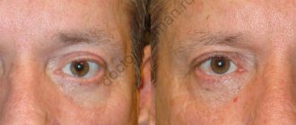
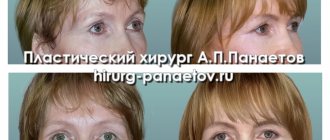
Rehabilitation after eyelid surgery with asymmetry correction: 6 days after surgery
Eyelid asymmetry is a pronounced unevenness in the position of the lower and upper eyelids or the left and right eyelids.
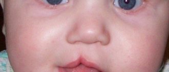
Different eye sizes in young children
Doctors state that the most harmless condition occurs when there is a difference in the size of the eyes in children from 3 to 5 years old. At this age, the formation of the muscular system occurs and slight asymmetry of the eyes actually occurs. However, even in such a situation, it would not be superfluous to consult an ophthalmologist, and in some cases, a neurologist. If the conclusion of these specialists is positive and the condition itself does not cause any particular concern, all that remains is to wait until the growing body corrects everything on its own, because such a condition does not require treatment.
Causes of eye asymmetry
Facial asymmetry can develop immediately after birth. This means that the condition began while still inside the womb. Another type of disease is pathological, and occurs several months or years after birth.
Anomalies of the skull structure
Incorrect skull structure is inherited. Defects may include various irregularities and depressions:
- the right side is larger than the left, or vice versa;
- the affected area is excessively large;
- sloping forehead or back of the head;
- one cheekbone is larger than the other.
All these factors change facial features, making them asymmetrical.
Developmental disorder of the lower jaw
The most harmless condition is malocclusion. A person develops a jaw that is pushed forward, a disruption in the normal development of wisdom teeth, and a receding or overly large chin. The lower jaw is one of the largest parts of the face, so its violations affect a person’s appearance.
Muscle and connective tissue problems
If a person has weakness in the facial muscles, drooping of the eyelids, cheeks, and eyebrows occurs. Reduced quality of connective tissue contributes to sagging skin. If this condition is common on one side of the face, it will be asymmetrically lower than the other.
A typical picture of muscle and connective tissue disorders is one eye wide open, the other narrow.
If the muscle tissue disorder can be not only on the face, but also on the neck. If it is very tense, the person's head will lean in this direction.
Violation of the structure of the temporomandibular region
If a person has a disorder in the joint between the lower jaw and the temporal bone, movement is blocked when opening the mouth. It is formed unevenly; a person can open first to the right side, then to the left, and vice versa. If the condition is severe enough, the person must first move it, then open it.
Strabismus
Strabismus develops in one or both eyes. Defects form immediately after birth or after some time. The causes are various neurological and ophthalmological primary diseases. The coordinated work of the eyes is disrupted. If one organ of vision decreases in acuity, it gradually turns off its function.
Vision defects
There are various diseases that reduce vision function. Many of them form external changes. A person develops a cataract, a cataract, or a scar on one eye. At the same time, the second eye remains healthy, this forms asymmetrical facial features.
Mechanical damage and inflammatory conditions of nervous tissue
Such diseases lead to swelling. If the nerve is inflamed on only one side, it increases sharply in size. Additionally, a pain syndrome is formed. This is observed with mechanical damage, the motor activity of one side is impaired. A person retains facial expressions only on the healthy side.
Absence of a large number of teeth
If the changes are only on one side, the cheek is pressed inward. The healthy part of the face remains normal. A person's face changes when talking. A lisp and a lack of pronunciation of certain sounds appear.
Bad habits
These include:
- squinting of one eye;
- Constantly chewing gum or foreign objects;
- sleep on one side.
Defects develop gradually. A bad habit affects muscle, connective and bone tissue. The location of muscles and bones relative to each other changes. If a large area is affected, the defect can only be corrected through surgery.
conclusions
So, let's draw the line. When a noticeable difference in eye size appears, you should definitely pay attention to possible additional symptoms. The appearance of swelling of the eyelid, redness of the mucous membrane and purulent discharge always indicate a bacterial infection. If the situation is aggravated by periodic paroxysmal pain, this is most likely neuralgia. But when the resulting difference in eye size is not accompanied by any symptoms, it is worth assuming a pathological process in the brain.
However, all the situations considered require the prompt intervention of a specialist. You need to remember this and not delay your visit to the doctor!
In the medical department, everyone can undergo examination using the most modern diagnostic equipment, and based on the results, receive advice from a highly qualified specialist. The clinic provides consultations to children from 4 years old. We are open seven days a week and work daily from 9 a.m. to 9 p.m. Our specialists will help identify the cause of vision loss and provide competent treatment for identified pathologies.
You can find out the cost of a particular procedure or make an appointment at the Moscow Eye Clinic by calling 8 (800) 777-38-81 (daily from 9:00 to 21:00, free for mobile phones and regions of the Russian Federation) or using the form online entries.
Yakovleva Yulia Valerievna
Treatment
There are various treatment methods. They are selected by the doctor individually for each patient. He, in turn, chooses the most appropriate method:
- Facial correction using cosmetics. If it is necessary to narrow the area, it is darkened. If you expand it, it brightens it up. Corrective foundation creams and pencils are used for this.
- Maxillofacial or plastic surgery. They contact a plastic surgeon, who individually selects the type of operation for the patient.
- Visiting the dentist. He can install crowns or new teeth.
- The use of gymnastics and massage complexes for reduced muscle tone.
- Treatment by a neurologist to eliminate the inflammatory condition of the nervous tissue.
Facial asymmetry can be minor or severe. In the latter case, patients want to change external parameters. To do this, they turn to doctors of various specialties: dentist, neurologist, ophthalmologist, plastic or maxillofacial surgeon. After proper treatment, facial features become symmetrical.
The essence of the operation
At the first stage, the surgeon applies markings to the skin of the eyelid. This stage of the operation is considered one of the most important, since it is the correct identification of the problem and ways to eliminate it that determines the final result.
Preoperative marking
At the next stage, the surgeon makes an incision and removes excess skin and fatty tissue.
Excision of excess skin and fat tissue
Cosmetic stitches are then placed on the incision. Since all manipulations are performed in the natural crease of the eyelid, the likelihood of postoperative scars is minimal.
Cosmetic stitches
Next, the suture line is sealed with a thin strip of a special adhesive plaster and the operation can be considered complete. As a rule, blepharoplasty is performed under local anesthesia. In this case, the patient does not experience any pain. The duration of the operation varies from 30 minutes to 2 hours.
Contraindications
- the reason for the formation of asymmetry is ophthalmological in nature;
- the eyelid has already blocked a third/half of the pupil or the entire pupil;
- oncology and HIV;
- bleeding disorders;
- uncompensated diabetes mellitus;
- acute diseases;
- diseases of the cardiovascular system;
- exacerbation of chronic pathologies;
- severe myopia;
- chronic eye diseases, dry eye syndrome, increased intraocular pressure.
Ptosis is not just a cosmetic defect
Of course, a cosmetic defect is much more noticeable than a functional defect, and therefore worries many patients more than the danger of a decrease or loss of visual functions. Meanwhile, almost any discrepancy between the real state of complex ocular mechanics and its regulatory status creates a direct threat to vision.
Thus, the most obvious danger is that a gradually drooping (or simultaneously drooping, as happens, for example, with strokes) eyelid creates an optical interference in the path of the light flux through the pupil to the retina. With partial or incomplete ptosis, a person is forced to take the so-called. “stargazer pose”: the head is thrown back, the eyebrows are raised, the forehead is tense and wrinkled, the facial expression is arrogant and sleepy.
With unilateral ptosis, especially in children, at the stage of ongoing formation of the visual system, such an insidious pathology as amblyopia, or “lazy eye syndrome” may latently develop: the signal from a partially blocked pupil, transmitted by the retina through the optic nerve, is perceived by the cerebral cortex as interference and over time is simply turned off and ignored; An eye that remains unused without its natural loads can become “lazy” and gradually degrade to the point of complete blindness. In some cases, due to chronic differences in eye tension, strabismus develops, visual fields narrow, etc.
The symptom complex typical for blepharoptosis also includes constant irritation and/or chronic inflammation of the eye (conjunctivitis, keratitis, combined keratoconjunctivitis), asthenopia (“eye weakness”, fatigue due to forced muscle strain), sometimes diplopia (double vision), dry eye syndrome, etc. . There are rare forms of ptosis with very specific phenomena. In particular, Marcus-Gunn syndrome is described, in which there is complex synkinesis - friendly, interdependent movements between the masticatory (chewing) muscles and the ocular levators (muscles that lift the eyelid upward).
A closed mouth is accompanied by blepharoptosis, but as soon as the patient opens his mouth, the eyelid rises; if you open your mouth even wider, the palpebral fissure expands proportionally. Despite the relative rarity and complexity of pathogenesis, this syndrome can be caused by a variety of unrelated reasons - from removal of the upper teeth and other mechanical injuries (TBI, damage to the facial nerve) to purely mental injuries.
Rehabilitation period
The patient can go home within 1 hour after surgery. The duration of rehabilitation depends on the volume of surgical intervention. The recovery period is 10-15 days, but the final result can be assessed only after 1.5-4 months. During the first few days, you need to provide complete rest to your eyes: avoid watching TV, reading books and working on a PC. If this is not possible, the use of special eye drops is indicated.
The sutures are removed 5 days after blepharoplasty; until this point, it is forbidden to touch the eyes with your hands; therefore, temporary refusal of contact lenses will be required. After removing the sutures, you are allowed to wash yourself carefully; treatment with non-aggressive antiseptics is indicated. For 2 weeks after surgery, you should refrain from physical activity, smoking, alcohol, strong coffee, makeup, salt, large amounts of liquid at night, and visiting swimming pools and spas. Excessively bright lighting should be avoided.