A mole is a pigmented formation on the skin of a benign nature. The scientific name of a mole is nevus, it can be congenital or acquired, and its color varies from light brown, red, purple to black. Depending on their location, birthmarks can appear on any part of the skin of the body or mucous membranes. The largest number of acquired birthmarks is found in people over 50 years of age. In children, moles begin to appear in the first years of life; in newborns, they are practically absent.
Moles appear due to the accumulation of pigment cells that are located between the dermis (inner layer of skin) and epidermis (outermost layer of skin). Moles can be located inside the dermis, on the surface or in the border layers of the skin. It can protrude on the surface or be located inside. In size, moles can be very tiny, or grow to gigantic sizes - about 10-15 cm. The greatest danger in terms of cancer degeneration are moles of large or very large sizes. The danger of any mole is that, under certain conditions, it can become malignant or develop into skin cancer - melanoma.
Main questions
- What moles can and should be removed?
- What do you need to know about diagnosing moles and other skin formations?
- What moles and neoplasms are considered “bad”? And what is “melanoma”?
Photo: kuprevich / freepik.com Elena Olegovna Belyanina, the leading dermato-oncologist at Frau Klinik, will answer these and other questions especially for readers of medportal.ru.
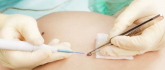
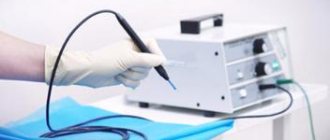
Cryodestruction as a solution for difficult cases
Mole removal can also be carried out using the cryodestruction method, that is, using liquid nitrogen. This method is quite controversial, since in addition to the visible advantages, it also has a number of significant disadvantages. Perhaps we need to start with the positive aspects. First of all, these include the absence of pain during the operation, a minimal number of consequences (the wounds heal painlessly), and the possibility of using the method to eliminate large formations. In addition, nitrogen acts as a kind of antiseptic and prevents bacteria from entering the wound.
Unfortunately, removal of a nevus with nitrogen often has to be carried out in several stages. So, it is not always possible to completely remove the formation the first time, and the manipulations have to be repeated. Among other things, the disadvantages include:
- The presence of a temporary burn on the skin;
- Negative effects on the body as a whole, and on the skin located around the nevus in particular;
- Long healing period;
- The operation is not performed on the face.
Prices for such an event range from 1000 to 1500 rubles. This accessibility makes cryodestruction quite popular.
Reason to see a doctor
— What neoplasms are considered “dangerous”? What should you pay attention to when new moles appear?
There is no exact standard by which a mole is considered “bad” or “good.” In each case, a thorough diagnosis is necessary. It’s great when people themselves come for preventive diagnostics of tumors. Every year in May, a charity event “Melanoma Day” is held, when you can get free consultation from a specialist.
The purpose of preventive examination of moles is not only to identify suspicious or malignant formations, but also to identify people with risk factors for the formation of melanoma (malignant skin tumor). Such patients should be under constant dynamic observation by a dermato-oncologist.
Photo: wavebreakmedia_micro / freepik.com
Important! The appearance of new moles after 40 years is a reason to contact a dermato-oncologist! When changing existing nevi, you should pay attention to such signs as rapid growth, changes in the color and structure of the neoplasm, compaction, ulceration and bleeding. In addition, people who have blood relatives with diagnosed skin melanoma are at risk!
Rehabilitation period
After the tumor is removed, it goes through several stages of healing.
- Immediately after laser treatment, the growth becomes covered with a crust. Under no circumstances should it be torn off or picked at. It should fall off on its own. This will happen within 7 days after the procedure.
- After the crust falls off, pink skin remains underneath. It should not be injured or exposed to direct sunlight. This period can last up to two weeks.
- At the last stage of healing, the formation of new tissue is completed and the redness completely disappears. If there is a small hole, it heals within a few weeks.
Diagnostics
— What methods of studying moles exist today?
Dermatoscopy of an atypical mole
Currently, it is impossible to imagine examining the skin without performing dermatoscopy. Dermatoscopy is a simple, non-invasive method for assessing the nature of skin tumors by visualizing the structures of the epidermis and dermis. When there is suspicion of a malignant nature of the formation, a biopsy is performed (removal of the tumor within healthy tissue with histological examination).
Histological examination (examination of tissue under a microscope) is the final stage of diagnosis. This study is also carried out when removing benign moles! It is necessary to understand that removal of a malignant tumor, mistakenly accepted as benign, without histological examination, will lead to late diagnosis and the impossibility of cure!
Rehabilitation
When removing small and medium-sized tumors, the patient can go home immediately after the procedure. Giant nevi are removed under anesthesia; after the operation you need to stay in the clinic for at least one day. As a rule, giant neoplasms are congenital; they are detected in 1% of 1000 newborns. It is recommended to start treating giant spots from 6 months of age.
Regardless of the size of the nevus, after its complete excision there are no relapses. If the tumor was not completely removed, it may resume growth. When a malignant nevus is removed without histology, adequate therapy and repeated excision of tissue in the area of the tumor, the oncological process can progress.
If you still have questions, you can ask them to the administrator by phone listed on the website or request a call back. You can immediately find out the approximate cost of the procedure. The final price will be determined after examination by a dermatologist. We warn you that administrators have general medical education and cannot always help with the details and subtleties of certain procedures. For a detailed consultation, make an appointment with a surgeon.
Primary source honey / September 2018
Treatment
— What methods of removing tumors exist today?
Indications for mole removal can be divided into medical and aesthetic. If the doctor did not reveal any suspicious formations during the examination, and the patient does not like some moles or are injured, then removal is carried out for aesthetic reasons, at the request of the patient. If suspicious formations are identified during examination, they are subject to mandatory removal with histological examination. In cases where a clearly malignant neoplasm is detected, the patient is sent for treatment and examination to a specialized institution.
Photo: kalinovskiy / freepik.com
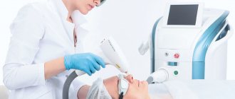
Today, there are several methods for removing tumors: surgical excision and destructive methods (radio wave, electrocoagulation, carbon dioxide laser). Benign tumors are predominantly removed using a laser due to the method being well tolerated by patients, the short duration of the procedure (3-5 minutes), and high aesthetic results.
Laser mole removal
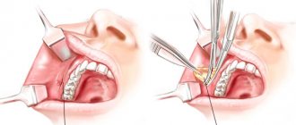
In certain cases, a surgical method is used (with suturing) - for large tumors, when located in skin folds, or if a malignant process is suspected!
How is laser mole removal performed?
Under the action of the laser, the neoplasm cells are heated and destroyed, and the laser also coagulates nearby blood vessels, so the risk of bleeding is negligible. During the treatment, the mole tissue and root are removed, which eliminates the risk of re-growth of the formation in this place. Depending on the size of the mole, removal is carried out over one or more procedures.
The operation is performed under local anesthesia, which eliminates pain. A small crust remains at the intervention site, which disappears on its own within 2 weeks. At this time, it is necessary to avoid moisture and friction at the site of manipulation, and also limit exposure to the sun; solarium, swimming pool, and bathhouse are also prohibited.
Should I remove a mole or not? Is there any danger?
— How justified are the myths that moles cannot be removed, that this leads to cancer and even death?
Moles can be removed, and sometimes it is necessary, but only after a thorough diagnosis. Undiagnosed (and therefore untreated) melanoma (malignant skin tumor) leads to death. Removing benign tumors does not in any way provoke the appearance of a malignant tumor.
Photo: wavebreakmedia_micro / freepik.com
Today there is a method that allows you to identify any malignant formation and make a decision on its further treatment. Dermatoscopy is a simple optical method for diagnosing and monitoring skin tumors. This method increases diagnostic accuracy by 45% (compared to naked eye examination)!
Biopsy and dermatoscopy of moles: we examine every “suspicious” nevus
Sign up for diagnostics of neoplasms at Frau Klinik: +7 +7 Clinic of Plastic Surgery of Professor Blokhin and Dr. Wulf metro station "Prospekt Mira" Moscow, st. Gilyarovsky, 55 metro station "Kurskaya", Moscow, Podsosensky lane, 20A
Recovery warning signs
Now that you know how to remove moles, you need to talk about in what cases you may need to see a specialist again. The recovery period after removal of a lesion is usually short-lived, however, if basic safety measures are not followed, certain problems may arise. So, see your doctor immediately if you:
- You feel severe pain in the area where the nevus was located;
- You notice a significant increase in body temperature;
- You feel a sharp, unpleasant odor from under the crust on the wound;
- You observe bleeding.
Knowing how to get rid of moles and how to care for the wound after removing the formations, you can become the owner of clear and beautiful skin.
Sources
- Patel M., Stuparich M., Nahas S. Total laparoscopic hysterectomy in combination with dilation and evacuation of an 18-week-sized uterus with gestational trophoblastic neoplasia: a novel treatment approach. // Am J Obstet Gynecol - 2021 - Vol224 - N3 - p.314-315; PMID:33197418
- Roelofs KA., O'Day R., Al Harby L., Hay G., Arora AK., Cohen VML., Sagoo MS., Damato BE. Detecting Progression of Melanocytic Choroidal Tumors by Sequential Imaging: Is Ultrasonography Necessary? // Cancers (Basel) - 2021 - Vol12 - N7 - p.; PMID:32664236
- Wen D., Ordonez D., McKenna A., Chang N.B. Fate and transport processes of nitrogen in biosorption activated media for stormwater treatment at varying field conditions of a roadside linear ditch. // Environ Res - 2021 - Vol181 - NNULL - p.108915; PMID:31759643
- Attia M., Hossny M., Zhou H., Nahavandi S., Asadi H., Yazdabadi A. Digital hair segmentation using hybrid convolutional and recurrent neural networks architecture. // Comput Methods Programs Biomed - 2021 - Vol177 - NNULL - p.17-30; PMID:31319945
- Monticelli D., Martina V., Mocchi R., Rauso R., Zerbinati U., Cipolla G., Zerbinati N. Chemical Characterization of Hydrogels Crosslinked with Polyethylene Glycol for Soft Tissue Augmentation. // Open Access Maced J Med Sci - 2021 - Vol7 - N7 - p.1077-1081; PMID:31049084
- Gale P. Using thermodynamic parameters to calibrate a mechanistic dose-response for infection of a host by a virus. // Microb Risk Anal - 2021 - Vol8 - NNULL - p.1-13; PMID:32289059
- Zhou X., Duan Z. Doppler ultrasonography reveals blood flow signals within the masses of invasive moles in subjects with normal hCG levels after chemotherapy: Three case reports. // Oncol Lett - 2013 - Vol6 - N4 - p.950-952; PMID:24137443
Alarms
These are the changes that should be especially alarming and serve as a reason for examination by an oncodermatologist and laser removal of moles:
- Changes in the size of the nevus, its rapid growth;
- The appearance of a red inflammatory rim around the nevus;
- Darkening or lightening of the nevus;
- The appearance of pain or itching in the area of the nevus;
- Changes in the surface of the nevus, the appearance of peeling, roughness, uneven contour;
- The appearance of another, daughter nevus on a nevus.
Moles that are located under a belt, under a collar, under a bra and in any other places that are subject to constant irritation should cause you increased attention.
Possible complications and side effects
The main disadvantage of surgical removal is the postoperative scar. It will be noticeable even with a cosmetic stitch, despite the fact that over time the skin will lighten and restore its structure. The surgeon’s task is to make the scar as invisible as possible, so choose a medical institution and doctor responsibly.
There is a risk of developing a keloid scar. This is a tumor-like growth of rough fibrous connective tissue. It is formed in people with a corresponding predisposition. Most often occurs when suturing large wounds.
Another risk is relapse. A repeat process is possible provided that the doctor has partially removed the mole, which is a gross violation of the surgical technique.
Immediately after excision, redness of the skin, itching, and a feeling of discomfort are possible. All these symptoms go away on their own after a few days. If something worries you, be sure to tell your doctor about it.