Cryodestruction of moles is the removal of moles from the surface of the skin using liquid nitrogen. The technique is based on the ability of liquid nitrogen to evaporate at a temperature of -196°C. Due to the ultra-low temperature, the liquid part of the cells crystallizes, and with subsequent thawing, the process of destruction of the nevus (mole) cells begins. In the capillaries that supply blood to the nevus, when frozen, thrombosis occurs, and the process of death of nevus cells intensifies.
Can all moles be removed using cryodestruction?
No, not all moles can be frozen with liquid nitrogen.
Nevus cells are formed in the skin during embryonic development, and moles can appear both at birth and throughout life. Nevi are of various origins and consist of pigment cells (melanocytes), connective tissue structures (vessels, collagen and elastic fibers), vascular tissue, and can be of a mixed nature. Most nevi are melanocytic, with varying depths of pigment. Some melanocytic nevi can become malignant and represent precursors to melanoma. This tumor is considered the most dangerous, as it is capable of rapid metastasis. Such nevi are called melanoma-dangerous.
Transformation into melanoma is possible in the following cases:
- excessive use of tanning, sunburn, especially in childhood;
- hereditary predisposition (presence of melanoma in relatives);
- hormonal surges during puberty, pregnancy, menopause;
- large size of pigmented nevi (more than 2 cm);
- growth in size, appearance of asymmetry, uneven coloring, compaction, bleeding of the pigmented nevus itself;
- having fair skin, blue eyes and red hair.
Dermatoscopy plays a major role in identifying melanoma-dangerous nevi - a hardware examination of the skin under 10x magnification with additional lighting. Externally, a mole may be small in size and have no signs of malignancy, but when performing dermatoscopy, the doctor may suspect the formation or risk of melanoma.
In such cases, the melanocytic nevus is removed using a surgical method (excision), capturing healthy tissue and then conducting a full histological examination. Experts do not recommend any other methods for removing melanocytic nevi.
Why are moles dangerous?
The ability to develop into a malignant state: to turn into cancer and melanoma depends on the type of mole. The most dangerous are dysplastic nevi, which are a variant of the development of ordinary melanocytic nevi (dermal, mixed and borderline), due to their transformation into melanoma. All melanocytic nevi are resistant to removal with liquid nitrogen. It is required to either freeze deeply, for a long time and several times. Or, don't freeze it at all. Another group of moles are non-melanocytic nevi (epidermal, sebaceous), seborrheic keratosis. They are capable of producing foci of squamous cell skin cancer and basal cell carcinoma. This group of moles can be removed very well and quickly with liquid nitrogen. In addition, nitrogen is used to treat foci of cancer in these moles, if they have just arisen (cancer in situ). It is extremely important that the doctor is clear about what he is removing during the procedure.
Sign up for a consultation
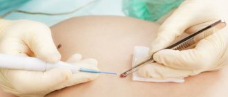
How is cryodestruction of moles performed?
Removal of a non-melanocytic nevus using nitrogen should be preceded by an examination by a dermatologist with mandatory dermatoscopy. Additional tests are usually not required. Mole removal is performed in two ways:
- contact method - using a cotton swab on a stick moistened with liquid nitrogen;
- in a non-contact way, when freezing occurs with a jet of liquid nitrogen aerosol, which is sprayed through a cryodestructor.
Anesthesia is possible, but is often not used, since the freezing procedure with liquid nitrogen lasts no more than 40 seconds. Unpleasant sensations in the form of tingling and burning sensations are possible, but quite tolerable. During the freezing process, the mole becomes covered with a white coating, then redness and slight swelling of the skin in the affected area appears, followed by the formation of a protective tight-fitting crust or bubble, which after a few days also transforms into a crust. The process of peeling off the crust occurs after 2–4 weeks. At the site of the removed mole, young light pink skin is formed, which subsequently evens out its color with the surrounding skin; no traces of removal remain.
Removal of tumors with liquid nitrogen: main advantages
The main advantage of this method is its low invasiveness. After the destruction of the tumor, a crust forms on the skin, under which the healing process is actively underway. Soon the crust falls off, leaving no trace. There is practically no bleeding or pain during this procedure; there is no need for special preparation or anesthesia.
The benefits of using liquid nitrogen include:
- rapidity;
- short recovery period;
- no need for special care of the treated skin area;
- affordable prices for cryodestruction procedures.
It is also important that various moles, papillomas or warts, regardless of their nature, can be treated equally effectively. Therefore, if you decide to remove a skin tumor by cryodestruction, you can count on a positive result after the first session.
How is the rehabilitation period after cryodestruction of moles?
In rare cases, on the first day after freezing with liquid nitrogen, there is discomfort in the form of soreness at the cryodestruction site. In this case, you can take 1 tablet of painkiller once.
After removing a mole with liquid nitrogen, it is important:
- do not injure the formed bubble or crust;
- Avoid contact of water, cosmetics, and detergents with the fireplace;
- do not rub with a washcloth, do not scratch the area of cryodestruction.
If redness and pain appear in the discharge area of cryodestruction, you should contact a specialist for a second appointment.
What complications can there be after cryodestruction?
The removal itself takes place quite quickly, but the process of tissue restoration takes much longer.
At the end of the procedure, the area of skin where the mole was is slightly sore and becomes hard. In rare cases, the patient may experience mild tingling and burning.
During the first 24 hours, the inflammatory process begins. Don't worry, this is normal. It’s just that the skin gets stressed after massive cell death. Then the place where the mole used to be begins to become covered with a crust (granulation tissue).
Already on the 10th day, the epithelization process begins, that is, new skin forms at the site of the lesion. This lasts no more than one and a half months. After this time, full-fledged skin will form at the site of mole removal.
Best materials of the month
- Coronaviruses: SARS-CoV-2 (COVID-19)
- Antibiotics for the prevention and treatment of COVID-19: how effective are they?
- The most common "office" diseases
- Does vodka kill coronavirus?
- How to stay alive on our roads?
As for side effects, it is possible that the patient will have a scar or scar at the site of removal. It depends on the size of the mole and the elasticity of the skin.
Skin care after removal
- During the first 24 hours, you should not wet the removal site;
- Treat the wound with Fukortsin solution or use Baneocin powder until the crust formed after removal comes off on its own;
- For men: do not shave in the area of removal for 3-4 days;
- For women: do not apply cream and decorative cosmetics in the area of removal until the crust comes off on its own;
- Before going outside, use sunscreen if the removal area is on exposed parts of the body.
All superficial neoplasms - papillomas, keratomas, xanthelasmas, etc., as well as small formations, are removed without scars or marks on the skin, which guarantees a high cosmetic effect. After removal of large formations, in addition to the above-mentioned superficial ones, traces of hypopigmentation or barely noticeable scars may remain.
Where to go for help to remove tumors?
If you find a tumor on your body, you should not leave it unattended. You should know that the papilloma virus is an infectious skin disease that requires professional treatment. The sooner you contact professional specialists, the faster they will help you get rid of any tumors and unpleasant factors associated with them.
You can contact our specialized clinic “VIVO CLINIC” for help by phone or directly on the website. We employ qualified specialists who use innovative diagnostic and treatment methods using European quality equipment. Each patient receives an individual approach and high service. You can make an appointment with a specialist at any convenient time.
Sign up for tumor removal on the website or by phone 8 (8162) 900-900.
Contraindications and restrictions for removal of skin lesions
- Suspicion of the malignant nature of the neoplasm during dermatoscopy;
- Pregnancy and breastfeeding;
- Diabetes mellitus, decompensated course;
- Inflammation in the area of the neoplasm;
- Herpes in the acute stage;
- Infectious disease and elevated body temperature;
- Taking anticoagulants;
- Tendency to form keloid scars.
The experience and high qualifications of our specialists, clear diagnostics, as well as work on very professional equipment allow us to ensure maximum efficiency and safety of services and procedures for the removal of benign and borderline skin formations.
Common myths and fears of patients
1. Can a mole grow back after cryodestruction?
Answer: If the procedure was carried out by an experienced specialist who strictly follows the technology, a new tumor will not appear in the treated area.
2. Removing a mole can lead to the formation of a malignant tumor.
Before proceeding with the removal of tumors using liquid nitrogen, the doctor prescribes the patient a laboratory examination of warts, moles, papillomas and other tumors using special equipment. Removal occurs only of benign moles that cannot cause the formation of a tumor and other unpleasant factors.
3. Removing papillomas using liquid nitrogen is a painful procedure.
As a rule, cryodestruction does not cause any pain in an adult; only a slight tingling sensation may be observed.
Children and adults with a high pain threshold can feel some pain.
Types of skin tumors
- Benign: moles (nevi), warts, papillomas, angiomas, fibromas, keratomas, xanthelasmas, etc. are present on the skin of the face and body of many people. They do not pose a direct threat to life and health.
- Borderline: senile and actinic keratomas, cutaneous horn, etc. Potentially capable of transforming into a malignant form. Requires close monitoring or removal.
- Malignant: melanoma, epithelioma, sarcoma, etc. They are life-threatening and require treatment by surgeons and oncologists in specialized oncology hospitals.
Causes and types of warts
Warts are skin growths that look like a hemispherical nodule, a dense skin growth or a soft papule. It is possible to hope that the wart will disappear on its own only in childhood, when such resolution is observed in approximately half of all cases. As for adults, the spontaneous disappearance of an epidermal growth is the exception rather than the rule.
Doctors say that the papilloma virus, which is present in the body of almost every person, is to blame. This virus “dormants” for some time, remaining in a latent state, and it becomes active at the most inopportune moment, when the protective function of the immune system decreases.
Typically, warts appear on the surface of the skin, as well as the mucous membranes of a person. The most common types of warts are:
- Genital;
- Flat;
- Plantar;
- Ordinary (or vulgar).
More often , warts form in women and children. This has nothing to do with the widespread stereotype that this phenomenon occurs due to uncleanliness or violations of personal hygiene rules. In fact, children and women are at risk only because their skin is thinner and more delicate. In children, for example, only three layers are distinguished in the epidermis, while in an adult there are five. Frequent microtraumas and violations of the integrity of the skin barrier, as well as the imperfection of the children's immune system, also provoke an increased risk of skin growths in patients of the younger age group.
The most common are common or, as they are also called, vulgar warts. Their favorite place is the fingers and hands. It often happens that when people see a wart that appears, they try to remove the skin defect themselves by tearing it off. However, under no circumstances should you do this. Thus, repeated self-infection of nearby skin areas occurs.
Many people think that getting rid of a wart can be done very simply by cutting it off with a blade or sharp knife. But this opinion is erroneous, since it is unlikely that it will be possible to completely remove the wart, but it is possible to cause its further increase in size. It is also important not to confuse a wart with a tumor and take all necessary measures in a timely manner. As mentioned above, a wart looks like a spherical papilla or nodule, its base is located deep in the skin.
Sometimes warts cause very unpleasant and painful sensations, making it difficult to carry out daily work. Warts are considered a cosmetic defect, and may also indicate an abnormal growth of tissue, which can undergo a process of malignant degeneration, leading to a precancerous or oncological condition, therefore, if you notice such an epidermal growth, you should seek help from an experienced doctor as soon as possible, who will tell you how to It is better to remove a wart on the body.
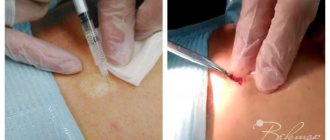
Preparation and carrying out the procedure
The appointment of a session is preceded by a consultation with a specialist. The doctor examines the tumors, selects the necessary laser parameters, and collects anamnesis to eliminate the risk of contraindications. If necessary, additional laboratory and instrumental studies are prescribed.
Before the procedure, the target area is decontaminated and treated with a local anesthetic. In combination with the laser cooling system, it provides complete patient comfort. Using the device's manipulator, the doctor acts on the affected skin in a targeted manner. High temperature burns damaged cells at a given depth, disinfects the wound surface and coagulates blood vessels. The whole procedure takes only a few minutes.
To completely remove a plantar wart, one session of laser treatment is usually sufficient. After treatment, the affected epidermis becomes covered with brown granulations, which disappear over time and leave clean, young skin.
Diagnosis of skin tumors
It is not always possible to visually determine the benignity or nature of a particular skin formation. Therefore, before removing any tumor, it is examined.
The main optical device used by a dermatologist to examine moles and other skin growths is a dermatoscope. The examination procedure using a dermatoscope is called dermatoscopy. This is a modern hardware diagnostic method that allows a professional experienced specialist to visually assess and diagnose the nature of any skin tumor under tens of times magnification.
Price for the procedure for removing skin tumors
| Dermatoscopy | 850 |
| Papillomas 1-2 mm (face and body) | 250 |
| Papilloma 1-2 mm (eyelid) | 650 |
| Xanthelasma up to 5 mm | 1500 |
| Keratoma 1-5 mm (face and scalp) | 650 |
| Wart 2-5 mm | 1950 |
| Moles up to 3 mm | 2500 |
| Moles larger than 3 mm | 4500 |
| Condylomas (single) | 850 |
A more detailed price list for procedures for removing benign skin tumors in our clinic is posted in the prices section.
What can be treated with liquid nitrogen?
This treatment method copes with warts and papillomas. Let's look at each category in order.
Removing warts with liquid nitrogen
Warts are growths that can form on any part of the body as an overgrowth of the top layer of skin. The size of a wart can be either 1 mm or several centimeters. From a medical point of view, such growths are benign formations that do not penetrate into surrounding tissues.
Warts can be:
- viral (most);
- venereal (sexually transmitted condylomas);
- vulgar (the effect of HPV on the skin area);
- teenage (with hormonal changes in the body);
- plantar (uncomfortable shoes or environmental influences);
- thread (excess weight, heredity, pregnancy);
- genital warts (sexually transmitted);
- seborrheic keratoma (senile warts that are not infectious in nature).
Removal of papillomas with liquid nitrogen.
Papillomas occur when the skin and mucous membranes are damaged by the human papillomavirus (HPV). This problem is the most common among the population. Most often it is transmitted to humans during sexual and standard contact with other objects or people through microtrauma to the skin. Activation of this virus occurs due to decreased immunity, which is closely related to problems such as:
- poor nutrition;
- oncological diseases;
- hormone therapy;
- unprotected sexual intercourse or promiscuity;
- stress and depressive disorders;
- gastrointestinal diseases;
- acquired immune deficiency syndrome.
The shape of the papilloma can be completely different, and the color is usually red, flesh-colored or brown.
What types of skin tumors are there?
The most common types of skin formations are:
- Moles. Pigmented neoplasms of various colors (black, brown, flesh-colored). They can be either flat or raised above the skin.
- Papillomas. Convex formations have a brown or dark tint. They differ in their viral nature and reduce immunity.
- Warts. Nodular or papillary-like projections on the skin. Usually caused by human papillomatosis viruses, they can be transmitted through contact with a sick person.
- Condylomas. A type of wart that occurs on the genitals. Transmitted sexually with the papillomatosis virus.
Removing each type of tumor is a responsible process that must be performed by professional specialists. Attempts on your own can lead to serious negative consequences for human health.
Treatment methods
Depending on the severity of the problem and individual contraindications, plantar tumors are removed in different ways:
- electrocoagulation with high-frequency current;
- cryodestruction with liquid nitrogen;
- surgery;
- conservative drug therapy;
- radio wave exposure;
- laser technique.
Laser removal is recognized as the most effective and safe in modern medicine. Light radiation burns damaged tissue without injuring nearby ones. At the same time, neighboring cells are “sterilized,” which eliminates the risk of re-formation of plantar warts. The non-invasive, non-contact method eliminates the possibility of infection, does not cause discomfort, and has a short rehabilitation period. Another advantage of the laser is a reduced list of contraindications. The technique is also used in patients with insufficient blood clotting, a tendency to allergies, weakened immunity, and impaired metabolism.
Laser removal of warts on the elbow. Wart removal in progress. No traces remain
Laser removal of warts on the finger. Removing warts on hands with laser. Fast and painless.
Laser removal of multiple warts on the hand. Laser removal of warts
Disadvantages of cryodestruction
Despite the advantages and popularity, this method also has disadvantages, due to which it is not recommended for use for removing moles on the face:
- Insufficient accuracy . The cryodestruction method does not allow you to control the depth and area of action, so during the operation the tissue surrounding the mole may be damaged.
- Lack of aesthetics . When removing large moles, scarring may form after healing.
- Possibility of mole regression . If the mole is not completely removed, regression is possible, which will lead to the need for repeated removal.
- Long rehabilitation . After the mole removal procedure, complete tissue healing takes much longer than using other methods.
In the Medicenter clinic network, moles are removed only using modern methods.