At the Center for Aesthetic Laser Medicine, moles are removed using a laser, so in the first hours after the procedure, a crust appears at the site of the removed tumor, and after 6–8 hours there is noticeable swelling and redness. This is a normal skin reaction to laser coagulation. On average, the wound healing process after mole removal can take from 2 to 4 weeks. It should be understood that the larger the area occupied by the removed mole, the longer it will take for tissue regeneration in the damaged area.
Healing stages and care
0–7 day
During the first 7 days after the procedure, a dark crust forms at the site of the mole, which under no circumstances should be removed. The postoperative crust on the wound performs a protective function - it protects the wound from infection and allows new tissue to actively grow underneath it. For the first week after removal, the operated area should be protected from all kinds of injuries and damage - avoid rubbing clothes or washcloths, do not comb, do not allow cream or other cosmetics to come into contact. If prescribed by a doctor, the crust can be treated with disinfectants (a weak solution of potassium permanganate) or ointments with antibiotics.
7–14 days
On days 7–9 after removal of the mole, the dark crust from the wound should fall off, leaving behind soft pink new skin. During this period and the next 10–12 days, the damaged area should not be exposed to ultraviolet rays. To do this, you should reduce your time in the sun and use sunscreen with a maximum sun protection factor of SPF 50. Direct exposure to the sun on young skin after removal of a nevus can lead to persistent pigmentation, which is especially undesirable on the face.
20 day and after
By the 20th day after the laser coagulation procedure, full-fledged healthy skin is formed at the site of the removed nevus, capable of resisting ultraviolet radiation and other external influences, including mechanical ones. After complete healing of the wound, no further specialized care is required. If a slight itch is felt at the site of the removed nevus after 20 days, but the skin looks normal, you can use special soothing ointments. By the 30th day after the procedure, the hole from the removed mole is smoothed out, after which, by the 3rd month, a barely noticeable trace of the procedure remains on the skin.
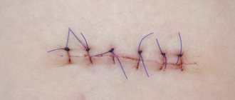
How does a wound heal?
The following phases of wound healing are distinguished:
- Inflammation. It begins immediately after the injury and lasts about 2-3 days, during this phase it is necessary to clean the wound and carry out anti-infective measures.
- Proliferation. From 3 days to 2 weeks. It is necessary to maintain the absence of infection and adequate hydration.
- The appearance of a scar. From 2 weeks to several years. During this phase, measures are taken to correct the scar depending on its type. [12]
General restrictions after mole removal
- For four weeks after the laser mole removal procedure, it is not recommended to visit the solarium or sunbathe in order to prevent hyperpigmentation;
- For two weeks after the procedure, it is recommended to refrain from visiting the sauna and swimming pool to reduce the risk of secondary infection;
- For 3–5 days after removal, it is necessary to avoid drinking alcohol, as they cause vasodilation, which can cause bleeding;
- If a mole was removed on the face, then it is not recommended to apply decorative cosmetics to the laser-treated skin for 7 days.
Which method is the most effective?
Unfortunately, there is no most effective treatment for scars. The most effective is considered to be a combination of various methods, which is always selected individually. Injections of corticosteroids into hypertrophic or keloid scars are considered an accessible, effective and inexpensive method.
Atrophic scars are distinguished separately: for their correction, planar ablative lasers, fractional ablative lasers (CO2 and Er:YAG), various combinations of lasers (fractional +PDL, Qsw+fractional lasers), collagen, fillers with hyaluronic acid, PRP therapy, microneedling are used. , peeling and a number of other methods [11, 12]
Previously, the dermabrasion method was used to correct atrophic scars, but now it is practically not used, since it is associated with the risk of side effects: bacterial or viral complications, telangiectasia, hyperpigmentation, a long rehabilitation period, as well as hypertrophic or keloid scars.
Complications after removal
After removing a mole, you must carefully monitor the damaged area of skin. In some cases, after the removal procedure, negative symptoms may be observed that require contacting the clinic and prescribing additional therapy. These symptoms include:
- purulent discharge from the wound;
- severe swelling of the skin that lasts for several days;
- prolonged bleeding from the wound;
- increased body temperature above normal;
- severe wetting, severe itching.
Bibliography:
- J Am Acad Dermatol. 2021 Apr; 74(4): 607–25; quiz 625–6. doi: 10.1016/j. jaad. 2015.08.070. Wound healing and treating wounds: Chronic wound care and management. Powers JG, Higham C, Broussard K, Phillips TJ.
- J Am Acad Dermatol. 2021 Apr; 74(4): 589–605; quiz 605–6. doi: 10.1016/j. jaad. 2015.08.068. Wound healing and treating wounds: Differential diagnosis and evaluation of chronic wounds. Morton LM, Phillips TJ.
- Ann Hum Genet. 2021 Feb 27. doi: 10.1111/ahg.12245. Gene-based evaluation of low-frequency variation and genetically-predicted gene expression impacting risk of keloid formation. Hellwege JN, Russell SB, Williams SM, Edwards TL, Velez Edwards DR.
- Int. J. Mol. Sci. 2021, 19(3), 711; doi: 10.3390/ijms19030711. Recent Understandings of Biology, Prophylaxis and Treatment Strategies for Hypertrophic Scars and Keloids. Ho Jun Lee, Yong Ju Jang.
- Br J Dermatol. 2011 Nov; 165(5): 934-42. doi: 10.1111/j.1365-2133.2011.10492.x. Laser and intense pulsed light therapy for the treatment of hypertrophic scars: a systematic review. Vrijman C, van Drooge AM, Limpens J, Bos JD, van der Veen JP, Spuls PI, Wolkerstorfer A.
- Laser Ther. 2013 Dec 30; 22(4): 255–260. doi: 10.5978/islsm.13-OR-20 Laser Scar Management Technique. Takafumi Ohshiro, Toshio Ohshiro, and Katsumi Sasaki
- Arch Dermatol. 2011 Sep;147(9):1108-10. doi: 10.1001/archdermatol.2011.248. A randomized split-scar study of intraoperative treatment of surgical wound edges to minimize scarring. Ozog DM, Moy RL.
- Plast Reconstr Surg. 2021 Mar;141(3):646-650. doi: 10.1097/PRS.0000000000004110. Effects of Botulinum Toxin on Improving Facial Surgical Scars: A Prospective, Split-Scar, Double-Blind, Randomized Controlled Trial. Hu L, Zou Y, Chang SJ, Qiu Y, Chen H, Gang M, Jin Y, Lin X.
- Curr Pharm Des. 2017;23(15):2268-2275. doi: 10.2174/1381612822666161025144434. The Effectiveness of Topical Anti-scarring Agents and a Novel Combined Process on Cutaneous Scar Management. Fang QQ, Chen CY, Zhang MX, Huang CL, Wang XW, Xu JH, Wu LH, Zhang LY, Tan WQ.
- Dermatol Surg. 2021 Jan; 43 Suppl 1:S3-S18. doi: 10.1097/DSS.0000000000000819. Keloids and Hypertrophic Scars: Pathophysiology, Classification, and Treatment. Berman B, Maderal A, Raphael B.
- Clinical and Experimental Dermatology, June 1, 2013. Combination of intense pulsed light and fractional CO2 laser treatments for patients with acne with inflammatory and scarring lesions. B. Wang, Y. Wu.
- Skinmed. 2021 Aug 1; 15(4):271-276. eCollection 2021. Techniques for Optimizing Surgical Scars, Part 1: Wound Healing and Depressed/Atrophic Scars. Konda S, Potter K, Ren VZ, Wang AL, Srinivasan A, Chilukuri S.
Possible consequences
In some cases, the healing process may occur with some deviations from the norm, which can be eliminated with additional procedures. Such deviations are the following situations:
- Recurrence of a nevus (reappearance of a mole) is possible if during the procedure not all cells of the mole were removed and some microscopic nevus particles remained in the same place. This situation does not pose any health risk and requires only observation. If a mole grows back, it can be removed again if desired;
- Hypopigmentation is the appearance of a white spot at the site of removal. This cosmetic defect is possible if the removed tumor is located in the deep layers of the skin. Also, sunbathing during the prohibited period can lead to the appearance of hypopigmentation. In any case, the resulting white spot does not require medical correction and will go away on its own in 1.5–2 years;
- A hypotrophic scar is a depression at the site of a removed tumor that occurs due to the low rate of tissue regeneration. A deep scar is an inconspicuous cosmetic defect and does not require any intervention, as it gradually smoothes out on its own;
- A hypertrophic scar is a bulge at the site of a coagulated mole, which may have a darker color than the surface of the skin. If the raised scar does not smooth out on its own after 3–6 months, then the doctor may prescribe drug therapy or additional cosmetic procedures to eliminate it.
Is a scar a reason to go to the doctor?
Reasons for visiting a doctor:
- aesthetic dissatisfaction;
- itching and pain in the scar area;
- the formation of deformities and contractures, which can lead to disability.
If scar tissue begins to develop pathologically and causes a keloid scar, it is better to consult a doctor.
There are factors that increase the risk of developing a scar:
- genetic predisposition;
- race (black patients are more likely to develop keloids);
- localization of injury (when removing tumors in the chest, neck and shoulders, the likelihood of keloid scars is higher);
- age (the formation of keloid scars is typical for young people)
Locations
Two thirds of relapses are metastases, one third are nodular formations in the area of the first operation. The location of the maternal tumor affects the fate of the patient:
- every third person operated on for melanoma of the scalp and neck develops tumors in the scar, and in women the problem occurs in the first year after treatment;
- transit metastases have the largest percentage in the group of melanomas of the extremity; this type of progression in women develops five years after excision, and in the male population - in the second and third years after surgery;
- with the initial lesion of the skin of the legs most often, in seven out of ten secondary tumors are found in the lymph nodes, in women in the next year, in men a little later;
- The primary process on the trunk in every fourth person is complicated by distant metastases.
Forecast
Every year, melanoma is diagnosed in an average of 15 people out of every hundred thousand adults, approximately 3 patients die, and with a fairly stable mortality rate in the last quarter century, men began to die more often. Gender determines a lot in the prognosis of the disease, other things being equal, but young people experience the disease with less difficulty.
The probability of death with a late relapse is many times lower than with an early relapse. Early detection of progression promises better treatment results.
It is extremely difficult to predict the course of melanoma, because even a common process is not considered absolutely fatal; this malignant disease does not often live up to expectations. Don’t guess “to be or not to be”, contact specialists if you have problems, or better yet, before they appear - we will always help.
| Read more about dermatological studies at Euroonko clinics | |
| Consultation with a dermatologist-oncologist | from 5100 rub. |
| Skin examination using the German FotoFinder device | 12100 rub. |
| Diagnosis of melanoma | from 5100 rub. |
Book a consultation 24 hours a day
+7+7+78
Bibliography
1. Semiletova Yu.V., Anisimov V.V., Vagner R.I. /Treatment of patients with primary skin melanoma. Current state of the problem // Siberian Journal of Oncology. 2010. No. 4. 2. Semiletova Yu.V., Anisimov V.V., Lemekhov V.G. et al./Risk factors for relapse after radical treatment of skin melanoma//Sibir.onko.zhur.; 2012. No. 2 (50) 3. Stroyakovsky D. L., Abramov M. E., Demidov L. V. et al. /Practical recommendations for drug treatment of skin melanoma // Malignant tumors: Practical recommendations RUSSCO #3s2, 2018 (volume 4. de Vries E., Bray FI, Coebergh JW, Parkin DM /Changing epidemiology of malignant cutaneous melanoma in Europe 1953–1997: rising trends in incidence and mortality but recent stabilizations in western Europe and decreases in Scandinavia// Int J Cancer 2003; 107. 5. MacKie RM, Bray C., Vestey J., et al./Melanoma incidence and mortality in Scotland 1979– 2003 // Br J Cancer 2007;96.
Why is relapse dangerous?
Relapse in melanoma is not only the appearance of a malignant formation in the scar, but also metastases. They most often affect the lymph nodes, lungs, skin, brain and liver. The possibility of removing a recurrent formation opens up prospects for a long life; if the operation is technically impossible, then the present and future will be occupied by drug therapy.
Survival for multiple organ metastases does not exceed six months; the results of drug therapy leave much to be desired, even with the use of innovative immuno-oncological drugs, which rarely promise more than two years of life.