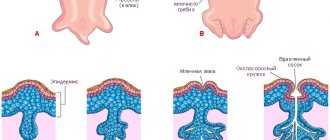
A gummy smile is characterized by a high position of the upper lip when smiling, when the gums above the upper teeth are noticeably exposed. Sometimes, when denoting a gummy smile, they speak of high gum level or a high smile. The condition and visibility of the gums plays a very important role in the perception of a person’s appearance. Ideally, the gums should not show much when smiling and should be in harmony with the cutting edge of the teeth. When aesthetically analyzing the appearance of the gums when smiling, it is necessary to pay attention to the degree of exposure of the gums above the teeth.
The normal position of the gums above the upper teeth when smiling:
From a physiological point of view, there is nothing wrong with a high smile with wide exposure of the gums! It just so happens all over the world that a smile without visualization of the gums or with very little visualization is considered more beautiful and harmonious.
Where does a gummy smile come from? A gummy smile is an anatomical structure. This is not a pathology or a deviation from the norm. It's just an anatomical feature, such as long fingers or short legs. With this physiology, the gums are exposed not only when a person smiles widely, but even during a conversation or sincere expression of emotions. Therefore, only the person who has such a feature should decide how to treat it: as a highlight or a defect that can be corrected.
The reasons for the change may be purely aesthetic. For example, when a man or woman, forming their personal image and working to improve their personality, come to the conclusion that a gummy smile does not look intelligent, sexy and charming enough, it simplifies the image, making the face rougher. It's always a personal feeling. The decision to correct a gummy smile must be made calmly, carefully, and in no case against the backdrop of emotions! And it is imperative to consult with a group of specialists - masters on various methods of correction. Why with a group of specialists? Yes, because there are several correction methods. To understand what is best for you, sometimes you need to listen to two or more specialists.
Dental method for correcting a gummy smile (surgical)
When a person has “a lot” of gum that opens above the upper teeth when smiling, then part of the gum above the teeth can be surgically removed. This is the most difficult correction method. Yes, the method is very effective! But the method is irreversible. Using surgery, you can achieve very good aesthetic results. The complexity of the method lies in the fact that there are surgical risks, i.e. healing of the gums and the formation of a new gum contour may not proceed as planned by the surgeon. Cutting off the gums above the teeth always leads to a visual lengthening of the teeth themselves. Those. The gums are shortened, and the teeth are lengthened due to the exposure of areas of the teeth that were under the gum. In some cases, this is corrected by aesthetic restorations on the teeth (veneers, restoration made from filling material). But in some cases, the visual lengthening of teeth after cutting off the gums itself leads to an aesthetic disaster! In order not to make a mistake, it is necessary to make the decision to trim the gums in order to correct a gummy smile not with one dental surgeon, but together with a surgeon and an aesthetic dentist. This specialist, after conducting a photo session of the patient’s smile, face and teeth, will evaluate the overall harmony. Only after this will it be clear whether the gum can be trimmed and how much gum to remove.
Orthopedic dentist S.V. Zukor. Dial-Dent specialist in correcting gummy smiles using dental methods.
Clinical case No. 1
The patient complained of excessive visualization of the gums, position of the teeth, and defective restorations.
Smile photo before:
Photo after gum surgery, stitches:
Photo after the stitches were removed:
Photo after completion of prosthetics (E-max veneers):
Photo of the smile after treatment:
Photos before and after treatment (comparison):
Cosmetological method for correcting a gummy smile (Botox)
There is an excellent method for correcting a gummy smile, which is used by Dial-Dent cosmetologists. The essence of the method comes down to the following. Botulinum toxin type A (for example, Botox or Dysport) is injected into the muscles surrounding the mouth and controlling the upper lip. Botox works by relaxing active muscles. After this, the person, even actively talking or smiling, simply cannot raise his lip as high as he did before. The effect of such a correction can be simply amazing! A bonus is an increase in the volume of the upper lip due to relaxation of the lip muscles.
The method is reversible. After 4-6 months, the effect of Botox wears off. This method of cosmetic correction also requires the highest qualifications and experience of a specialist! It is important to prevent asymmetry and choose the correct dosage and volume of the administered drug! And now a little secret! It is not necessary to use a lot of Botox to achieve excellent correction results. Dial-Dent specialists achieve results using only 8-15 units of Botox.
The cost of correcting a gummy smile using a cosmetic method ranges from 5,000 rubles (uncomplicated situation) to 9,000 rubles (complicated situation). Botulinum toxin is paid separately at cost. Botox - 150 rubles per unit, Dysport - 50 rubles per unit. Typically 8-15 units are needed. A good master achieves results by using a minimum of units of botulinum toxin, because a good master calculates the dosage more accurately, feels the anatomy of the muscle better, and more accurately calculates the injection site.
Cosmetologist Pavel Olegovich Kusin – specialist in cosmetological methods for correcting gummy smiles
.
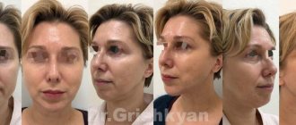
An example of gummy smile correction from the practice of specialists:
The girl underwent bite correction and teeth straightening. The smile has become more aesthetically pleasing, but a gummy smile, according to the patient, spoils the appearance. Cosmetological correction of a gummy smile was applied.
In this case, the cosmetologist introduced only 8 units of Botox. The result will be stable for six months. After this, if necessary, the procedure can be repeated. Repeating such procedures under the supervision of a good specialist is safe and harmless.
When correction is needed
Shyness, uncertainty, critical assessment of attractiveness and other negative mental states have a negative impact on social interaction. They can also cause significant difficulties when communicating, establishing work and personal contacts. In all cases when a gummy smile interferes with the harmonious development and formation of personality, or has a negative impact on any aspects of life, it is recommended to consult a dentist and consider correction methods to restore confidence in beauty.
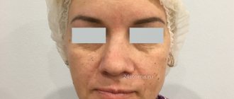
Gummy smile photo.
Speech therapy methods for correcting a gummy smile
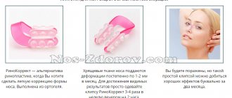
There are little-known methods for correcting a gummy smile with the help of a speech therapist-psychologist. There are only a few speech therapists who know these methods. Lips, lip mobility, tone of the muscles surrounding the mouth, and much more - all this relates to the human speech apparatus. A speech therapist is exactly the specialist who works with the speech apparatus and knows everything about it! The Dial-Dent speech therapist, using proprietary techniques and performing certain exercises with the patient in front of a mirror, teaches the patient to control the position of the upper lip and stop it when moving upward in the desired position. A speech therapist is needed only in the initial stages of this type of gummy smile correction. In the future, it all comes down to self-control. Over time, a person learns to control the degree of lip mobility, preventing it from rising high! The method is inexpensive! Absolutely safe! But it requires constant tension associated with self-control.
Speech therapist-psychologist "Dial-Dent" T.B. Zukor. Specialist in the field of non-medical and non-surgical correction of gummy smiles.
What is a gummy smile?
According to statistics, about 10 - 15% of all people on earth have a so-called gummy smile, in English it sounds like a “gummy smile”. What does she look like? It is customary to call a gummy smile a smile in which more than two millimeters of the upper gum is visible. Exposing less than two millimeters of the upper gum when smiling is considered normal. But the ideal smile would be one in which the gums are not visible at all, and only the upper incisors are exposed, while the lower front teeth are either not visible at all or only slightly visible.
Combination of all methods or several methods
In some cases, it is difficult for the patient to choose which path to correct a gummy smile to take. Separate practitioners each incline the patient to their own technique. By consulting with Family Dental, you have a huge advantage! In one institution we have gathered specialists in all known methods of gummy smile correction. You can listen to everyone's opinion at the consultation. If a decision is made to use one of the correction methods, or if a combination of methods is suddenly needed, we at Dial-Dent will always cope with the task perfectly.
Make an appointment by phone +7-499-110-18-02 or through the form on the website. You can ask questions about correcting a gummy smile to the chief physician of the clinic, Sergei Vladimirovich Tsukor, at
Gummy smile and short teeth syndrome: etiopathogenesis, classification, diagnostic criteria
Smile line assessment is one of the stages of the comprehensive aesthetic rehabilitation process. A gummy smile (GBS) is a non-pathological condition that causes aesthetic imbalance and is manifested by the exposure of more than 3 mm of the gingival profile when smiling (photo 1).
Photo 1. Patient with a gummy smile.
The reasons for the development of a gummy smile can be excessive growth of the upper jaw, too short an upper lip, or non-physiological eruption of the upper front teeth. There is also such a thing as short teeth syndrome (STS), which provokes excessive exposure of the gums or too short a tooth profile when smiling. Authors often say that DS and SCZ are related etio-anatomically and pathologically, and the diagnosis of one of the disorders should automatically make the doctor think about the presence of the second. The reasons for the development of DC can also be deep bite, excessive teething, compensatory teething (with pathological abrasion or erosion), gingival hyperplasia and other disorders of the processes of active/passive teething. The purpose of this article is to acquaint the reader with the classification of DS and the main methods of treating this condition, which will achieve the predicted results (photo 2).
Photo 2. View of the patient’s smile after treatment.
Researchers have previously proposed certain classifications of DD, but they related to the presence of this disorder in young people. The cause of DD may be changes in one of the following components of the dentofacial apparatus: the upper jaw, lip, gums, teeth, or in some of them at once. It is very important to determine the primary cause of the development of DU, since the entire subsequent treatment plan will depend on this. The authors of this article propose to use the modified classification of Monaco and colleagues as an adapted one. As diagnostic assessment methods, the doctor is recommended to analyze the medical history (taking into account age and general somatic health status), facial aesthetics, static and dynamic lip position, tooth height and incisal edge position, periodontal condition (probing depth, changes in the level of clinical attachment, height bone relative to the cemento-enamel junction). The essence of complex diagnostics is to connect visualized aesthetic changes in the smile profile with existing disorders in the oral cavity and the body as a whole.
Diagnostic process
1. History
The main aspects of the anamnesis analysis are the components of age and general somatic health status. The patient's age is important in assessing the associated stage of teething, and changes in general health, such as during pregnancy or taking certain medications, can help the doctor determine the cause of gingival hyperplasia (Figure 3).
Photo 3. Gummy type of smile in a patient with gingival hyperplasia.
2. Facial analysis
Assessing the proportionality of the face in its three anatomical zones from the frontal and lateral sides allows us to determine whether the patient needs changes in the area of the lower and middle third. An increase in the size of the middle third of the face may indicate the presence of an excessive trochanteric component of the maxilla. It is necessary to understand that the clinical assessment of the proportionality of the face is only approximate; for a more detailed analysis, an x-ray examination is required (photo 4). The increase in the size of the maxilla can also be recorded using cephalometric analysis, measuring the distances between the incisal edge of the central incisors and the plane formed by the anterior and posterior nasal spine (SPA-SPP) (palatal plane-incisal edge: 29-31 mm). However, according to the authors, these indicators are not accurate enough, given that the distance being studied may change depending on the degree of tooth wear, or on the amount of excessive eruption. For greater objectification, it is recommended to measure the distance from the palatal plane to the level of the cemento-enamel boundary, taking into account the fact that the position of this boundary changes depending on the effect of compensatory super-eruption.
Photo 4. Lateral radiograph of a patient with excessive vertical parameters of the upper jaw.
3. Analysis of the position of the lips and the condition of the perioral muscles
Analysis of the position of the upper lip in statics and dynamics (photos 5-6) allows you to diagnose the condition of a gummy smile. In a static position, the distance between the subnasal point and the lower edge of the upper lip is measured. The height of the skin part of the lip should be 15-16 mm, and the red mucous membrane should be 5-6 mm. With lower values, we can talk about the upper lip being too short. With a dynamic analysis, it is possible to diagnose hypermobility of the muscles of the levator labii superioris, which may be the cause of the development of the DU condition.
Photo 5. Measuring lip parameters.
Photo 6. Measuring lip parameters.
4. Analysis of the gingival profile
During the diagnosis, it is necessary to determine whether during a smile only the gum area in the frontal area is exposed, or whether the profile of mucosal exposure expands to the lateral areas (photo 7-8). In cases where DU provokes gum exposure only frontally, correction of this condition can be carried out through minimally invasive interventions, but when visualizing a wider gingival profile, it is simply impossible to do without an integrated approach to treatment to form an aesthetic contour.
Photo 7. Anterior gummy smile.
Photo 8. Anterior-posterior gummy smile.
5. Dental analysis
When diagnosing DU, the doctor must conduct a three-dimensional analysis of the position of the incisors in the structure of the facial profile. To do this, he asked the patient to pronounce the sound “m”, after which the lips took a resting position. The space between the lips in this condition should expose the profile of the incisal edge of the teeth by 0-4 mm, depending on the age of the patient. If the incisal edge is exposed excessively when the lips are at rest, the doctor may suspect that the patient has an oversized upper jaw, or a too short upper lip. If the incisal edges are insufficiently exposed, the condition of the teeth should be checked for symptoms of pathological abrasion. The latter are assessed relative to the average height and width of teeth described in the literature. In addition, it is important to understand whether the reduction in the vertical parameters of the teeth is caused by the effect of excessive abrasion, or by disturbances in eruption, or both. In addition, by analyzing the condition of the incisal edge, the clinician can determine the etiology of the disorder, whether it is associated with the tooth itself, or with the gums surrounding it.
6. Periodontal analysis
During periodontal analysis, it is necessary to determine the presence or absence of pathological and non-pathological disorders in the periodontal structure. A periodontal probe is used to change the depth of the pockets, the level of clinical attachment and the size of recessions. In cases where the patient has short teeth without signs of abrasion, an assessment of the periodontal condition is carried out in order to register possible inflammation, mucosal hyperplasia and eruption disorders. Eruption disorders are clinical situations in which the level of the gums reaches the surface of the enamel, which provokes a visual shortening of the crown. It is necessary to clearly understand the difference between active eruption disorders and passive eruption disorders, since these two conditions require completely different treatment approaches. Diagnosis of the area of the cement-enamel border is one of the methods for assessing the excessive size of the gums relative to the size of the clinical crown. This manipulation is carried out using a periodontal probe or dental probe No. 17.
The authors of this article developed a method for radiographic evaluation of eruption disorders (AltErX), which involves taking radiography after fixation of orthodontic wire at the gingival level with a flowable composite. In this way, it is possible to visualize the difference between the clinical and anatomical crowns (photo 9-10). When performing radiography, it is worth placing the radiographer as perpendicular to the tooth as possible, since distortions in the radiographic images can cause false results in patients with a very high or low palate. If eruption disorders are suspected, the doctor should also assess the intactness and level of the surrounding bone tissue, having previously performed an anesthesia procedure. This manipulation is mandatory before performing any periodontal interventions.
Photo 9. Fixation of orthodontic wire in the area of the gingival margin for the purpose of further radiological control of the relationship between the position of the cemento-enamel border and the level of the gums.
Photo 10. Fixation of orthodontic wire in the area of the gingival margin for the purpose of further radiological control of the relationship between the position of the cemento-enamel border and the level of the gums.
Discussion
The proposed modified classification, taking into account the etiology of DS, allows the doctor to have a more reasoned approach to the choice of treatment method for this condition. Most doctors agree that the main cause of DU is excessive vertical growth of the upper jaw. This is most often observed in patients without existing stable centric relation and centric occlusion positions (as in patients with a class 2 bite), with too deep a bite, and in the presence of a step between the incisal plane in the frontal region and the occlusal plane in the distal region. In the conditions described above, the dentofacial apparatus tries to compensate for vertical changes in occlusion through compensatory teething. When a gummy smile develops due to excessive wear of teeth and changes in the proportions between the pink and white components, experts speak of an acquired form of the disorder. In fact, it is caused by the fact that when smiling, a normal amount of gum is exposed, but since the crown height has decreased, the mucosal profile is dominant within the visualization limits. A gummy smile may also be associated with the height and position of the upper lip, as well as muscle hyperresponsiveness.
Thus, DU, on the one hand, can be caused by excessive visualization of the gums in the normal state of the teeth or changes in the parameters of the teeth themselves in the normal profile of the mucous membrane. Changes in dental parameters can be caused by pathological compressibility, attrition and erosion. Often such conditions are observed in bruxists and clenchers. In addition, disruption of the functional patterns of occlusion itself can cause the formation of an irregular contour of the incisal edges. Gum hyperplasia, in turn, can be caused by inadequate patient care for the condition of the oral cavity, or by the action of certain medications.
To select a method for correcting a gummy smile in conditions of intact incisal edges of the teeth, the doctor must perform a bone crest probing procedure and x-ray diagnostics to validate the level of bone tissue relative to the cemento-enamel boundary. In addition, any manipulations should be carried out taking into account the parameters of the general profile of the face and the patient’s physiological stage of eruption of problematic teeth. In order to diagnose changes in the position of the gums relative to the cemento-enamel junction, the authors recommend using a modified technique with a radiological marker fixed along the gum line. CBCT data can also be used for the same purpose. As an alternative, Cairo et al proposed a method for diagnosing tooth eruption disorders by comparing the parameters of the clinical crown of the tooth with the results of its radiographic diagnosis. There are rather contradictory data in the literature about what level of gums and teeth should be visualized when smiling. Indeed, even with exposure in some cases of more than 4 mm of mucous membrane, some types of smiles remain quite attractive, and, on the other hand, in certain situations, even minimal exposure of the gum profile leads to a complete aesthetic imbalance. The effect of a gummy smile in some patients can be compensated by the position of the lips, the adequate proportion of the size of the teeth and the appropriate condition of the muscles.
Conclusion
When treating patients with a gummy smile or short tooth syndrome, the first thing the doctor must do is to conduct an adequate diagnosis with an analysis of all possible etio-anatomical-pathological relationships. Sometimes the above-mentioned conditions are associated not with one, but with several etiological factors at once, therefore, their treatment should be carried out comprehensively. With proper preliminary diagnosis, it is much easier for the doctor to plan a rehabilitation protocol, ensuring the stabilization of the white and pink components of the smile with the formation of its proper and harmonious profile.
Authors: Antonello F. Pavone, DDS Marjan Ghassemian, BDS, DDS Simone Verardi, DDS, MSD
Botulinum therapy: features and indications for use
Botulinum therapy in Krasnogorsk today is an absolutely safe procedure that allows you to fight expression lines around the eyes, on the forehead and between the eyebrows. Professional cosmetologists also recommend botulinum toxin injections to correct the area around the lips, neck and eyebrows. The introduction of a microdose of botulinum toxin relaxes and temporarily paralyzes the muscle, as a result of which the skin is evened out and facial wrinkles are smoothed out, while maintaining natural facial expressions and freshness of the skin.
Xeomin, Dysport, Botox, Botulax are all trademarks of botulinum toxin.
Should a child with a gummy smile be taken to the dentist?
A visit to the doctor is necessary. It is important to find out the cause of the deviation and understand whether the child needs correction. Pediatric dentistry in Moscow has sufficient capabilities to predict the possibility of functional disorders occurring in the future. At an early age, detected anomalies are corrected much easier and faster. And aesthetic braces for teenagers have even become a fashion trend lately.
If the health of the heir is not in danger and there is no need for treatment, parents should not focus on the existing deviation. Otherwise, already in adolescence, a slight aesthetic defect can develop into a serious psychological problem.
Causes of gummy smiles in children and adults
In adults, a gummy smile is quite rare, since this problem in most cases appears in childhood. There are several known main reasons for the appearance of this problem: improper development of the facial skeleton, a short upper lip that cannot cover the gum, or, on the contrary, an overly large gum that covers the upper row of teeth more than necessary. At the same time, a strongly pronounced gummy smile, as a rule, is a consequence of malocclusion, or anomalies in the development of the maxillofacial skeleton.