The mammary gland consists of approximately 10 - 12 lobes. All of them should normally be grouped together in the chest area. An accessory lobule of the mammary gland is an atypical location of part of the glandular tissue of the organ. A woman may not know about its presence for many years, since it is not always accompanied by any clinical signs. What are the dangers of an additional lobe of the mammary gland and does it need to be removed?
Reasons for the appearance of an additional lobe of the mammary gland
It is necessary to distinguish between the concepts of “additional lobule” and “gland”. The pathologies are somewhat similar, but there are significant differences. The accessory breast (polymastia), in addition to glandular tissue, has a direct excretory duct, its own areolar zone and nipple. This is like a full-fledged mammary gland, only in a rudimentary size.
An additional lobule is just a piece of glandular tissue that, due to some circumstances, has ectopized into another zone. Its duct opens into a common one for all on the main chest.
No reliable reasons for the formation of an additional lobe of the mammary gland have been established. The generally accepted opinion is that this occurs even during the period of embryonic development under the influence of some unfavorable factors. As a result, some of the cells remain in the armpit area, and all the rest migrate to the usual location of the breast.
This can be either the influence of harmful environmental factors, or medications in the early stages of pregnancy, infectious diseases during this period, etc.
A hereditary predisposition to this feature has been noted, so it is possible that the pathology is associated with mutations of some specific genes.
We recommend reading the article about lymph nodes in mastopathy. From it you will learn about the causes and symptoms of pathology, diagnostics and treatment methods.
And here is more information about lobular breast cancer.
Diagnosis of the causes of enlarged axillary lymph nodes
- Consultation with a mammologist. The mammologist at the Clinical Hospital on Yauza will conduct a visual examination of the mammary gland, palpation, ask the patient about when the lymph nodes were enlarged, whether this is associated with a period of exacerbation of any disease, whether the patient had contact with infectious patients, etc. Next, the doctor will prescribe a comprehensive examination.
- Instrumental studies:
- Ultrasound of the mammary glands;
- ductography;
- digital and MR mammography.
Disruption of the lymph nodes can cause an increase in the accumulation of toxic substances by up to 83%. As a result, the kidneys, liver and other organs will suffer. Book an appointment with a doctor to eliminate malfunctions in the body and prevent the development of serious pathologies.
Symptoms of an additional lobule under the arm
Small additional lobules may not show themselves for a long time. Typically, a woman becomes aware of their presence under the following circumstances:
- During pregnancy, sometimes even before a missed period. This is due to the fact that under the influence of prolactin, the mammary glands are transformed for subsequent lactation. Moreover, all these changes occur in the additional lobule. Swelling appears in the armpit area, possibly even discomfort or pain.
- After childbirth, especially if the girl is supporting lactation. On the 3rd - 5th day, the maximum milk supply normally occurs. At the same time, the breast increases significantly, becomes dense and sensitive to the touch. Everything proceeds similarly in the additional lobe.
Typically, 3–5 days after childbirth, women notice pain in the axillary region and upon palpation a tumor-like formation is detected. There is no need to do anything in such situations, everything will go away on its own in 2 - 3 days. In rare cases, if the outflow of secretions from the additional lobule is disrupted, inflammation may develop in it - lactostasis and mastitis, with an increase in temperature and a deterioration in the general condition. Treatment follows the general rules for this pathology.
- If an inflammatory process occurs in the accessory lobule or other diseases (even cancer). Lactostasis can occur at any time during breastfeeding, and non-lactation mastitis of the accessory lobules (inflammation at any age) also occurs. In this case, a woman may think that her lymph node has become inflamed, a boil has appeared, or something similar. With the same frequency as in a normal mammary gland, a cancerous tumor can arise in an additional lobule. Typically, a woman discovers a small formation in her armpit, painless, mobile or not relative to other tissues. Therefore, it is important to conduct research in this area during mammography or ultrasound.
- With the development of signs of mastopathy. The disease can occur at any age in different forms. The main symptoms are the appearance of soreness in the armpit area in the second phase of the cycle, everything goes away with the onset of menstruation. Sometimes even the appearance of a small compaction is detected, which regularly disappears and appears again.
It is in these conditions that the mammary glands undergo serious changes, and the additional lobule “gives” itself.
So, you can suspect pathology if you have the following symptoms:
- the appearance of a compaction in the axillary region, less often it is the subclavian and others;
- formation is painful or not;
- Almost always the occurrence of symptoms is associated with pregnancy, childbirth, and the menstrual cycle.
Sectoral breast resection
Table of contents
- What is sectoral resection?
- Indications for surgery
- Contraindications for carrying out
- Is it possible to avoid sectoral resection of the gland?
- Preparation
- Operation progress
- What are the complications of sectoral resection?
- Does breast deformation remain after sectoral resection?
- Recovery period
- Advantages of carrying out the procedure at MEDSI
Modern surgical methods for treating glandular tumors are focused on preserving the operated organ and its functions.
- Resection is removal
- Sector - a triangular area with an apex at the nipple, constituting no more than 12–13% of the gland
Sectoral resection of the mammary gland is a precise surgery that allows you to remove a local tumor without affecting healthy tissue. Such operations are performed using aesthetic surgery methods. Breast deformation after surgery is minimal, the stitches are cosmetic. Minimally invasiveness allows manipulations to be performed under local anesthesia and significantly reduces the rehabilitation period.
Intervention is possible for small tumors in the early stages of the disease. Both formations of a malignant nature, which is confirmed by a biopsy, and benign ones, capable of degeneration, as well as foci of chronic inflammation with irreversible tissue deformation are subject to removal.
The method is also used for primary examination of tumor tissue.
What is sectoral resection?
Lumpectomy or sectoral resection of the mammary gland is the removal of the affected segment for therapeutic and diagnostic purposes. As a result of the operation, a tumor up to 3 cm in diameter with underlying tissues is isolated and selectively removed (at least 1 cm around the tumor to prevent relapses). The tumor is sent for histological examination.
At the same time, one or more lymph nodes involved in the process are removed - if they increase in size, which makes it possible to suspect the presence of metastases in the lymphatic system. Lymph node tissue is also sent for analysis.
The method allows you to preserve most of the glands, which remain capable of natural functioning - after the operation, the woman is able to breastfeed.
Indications for surgery
Indications for surgery are the presence of confirmed fibroadenoma or adenocarcinoma of the breast. To do this, the patient undergoes a series of sequential tests, which are prescribed to him by the attending gynecologist or mammologist:
- Ultrasound or sonoelastography of the mammary glands
- Mammography or computed tomography with contrast - the introduction of a substance that accumulates in atypical tumor tissue and makes the tumor brighter in the images
- Puncture (biopsy) - removal of a column of tissue from a suspicious neoplasm with a special needle for further examination of the atypicality of its constituent cells
The following are subject to removal:
- Rapidly growing benign glandular nodules ( fibroadenomas
) - Adenocarcinomas
at an early stage - Intraductal papillomas
, also capable of degeneration and creating the preconditions for lactostasis (milk retention) in women planning pregnancy - Lipomas
are pathological growths of adipose tissue localized within one segment or single - Granulomas
are encapsulated foci of inflammation - Foci of purulent melting of tissue, purulent infiltrates
that cannot be treated or restored
Contraindications for carrying out
- The location of the lesion in the deep layers of tissue
- The size of the formation is more than 3 cm
- Systemic diseases of the blood and other connective tissues
- Diabetes
- Presence of other cancers
- Pregnancy and lactation
- Acute infectious process
Is it possible to avoid sectoral resection of the gland?
At the stage of diagnosing a neoplasm, if its location allows, it is permissible to replace the excision of the lesion using sectoral breast resection with a biopsy, however, a biopsy does not completely remove the tumor, and if its malignant nature is confirmed, surgery cannot be avoided.
Growing fibroadenomas are also subject to mandatory removal, since they do not respond to conservative therapy and do not resolve on their own, and in addition, they tend to undergo rather rapid malignancy (malignancy).
The same applies to purulent abscesses that cannot be treated with medications - the presence of purulent infiltrates in the tissues, if left untreated, sooner or later leads to sepsis (a generalized bacterial infection of the blood).
Compared with other methods of radical treatment, sectoral resection of the gland has a number of advantages:
- Minimally invasive – preserving the functions of the mammary gland and the appearance of the patient
- Low risk of complications both during surgery and in the postoperative period
- Fast recovery – discharge is carried out on the third day after the procedure
Therefore, the possibility of performing such an operation for surgical diseases is considered as an advantage for the patient.
Preparation
Before a planned hospitalization for surgery, the patient needs to collect a number of up-to-date information about his condition. The absence of blood infections, the state of the hemostasis system, hormonal levels, heart function, and thyroid function are assessed.
The surgeon must provide:
- Blood tests: Coagulogram (prothrombin index, INR, fibrinogen, APTT)
- Blood type, Rh
- Thyroid stimulating hormone (TSH) level
- Female sex hormones (estrogens, gestagens, testosterone, prolactin)
- Blood biochemistry (ALAT, ASAT, urea, creatinine, bilirubin, glucose)
- Markers for hepatitis and HIV
- Electrocardiography
Operation progress
Depending on the type of anesthesia (general anesthesia or local), the patient is prohibited from eating for 12 or 4 hours before the procedure. You can drink water for the last time 3 hours before the intervention.
- The patient is laid on his back on the couch, under the control of mammography, the surgeon marks the surgical field; the duration of the operation and its effectiveness will depend on the accuracy
- The patient is then anesthetized and covered with sterile surgical drapes, leaving only the surgical site exposed. As a rule, premedication is given before anesthesia to reduce anxiety levels.
- Semi-oval incisions along marking lines from the periphery to the center (from the edge of the gland to the areola) end three centimeters from the location of the tumor
- Soft tissue is separated from the muscular fascia of the pectoralis major muscle using a blunt instrument
- The tumor is fixed in the surgeon's fingers and excised
- Tissue is removed within a radius of 10 mm around the tumor
- Vessels are coagulated or sutured
- Drainage is installed to drain blood and lymph
- The wound is then either sutured or covered with sterile material until the results of histological examination are obtained.
- A sterile bandage is applied over the suture, which is then regularly changed for dressings.
- The patient is removed from anesthesia and transported to the ward, where he remains under observation for 3 days
What are the complications of sectoral resection?
- Hematoma
(accumulation of blood in the wound cavity and nearby tissues) - eliminated with repeated intervention by suturing the vessel, administering hemostatic drugs - Seroma
(swelling due to lymph accumulation) – is removed using a puncture, the resulting fluid is sent for examination to exclude infectious complications - Infection
(entry of pyogenic flora into the wound with the subsequent development of purulent-septic complications) - antibacterial therapy is carried out - Lymphatic edema
(arms and neck from the side of the operated gland) - occurs as a result of resection of not one, but a whole group of lymph nodes during a malignant process; requires long-term specific treatment
Does breast deformation remain after sectoral resection?
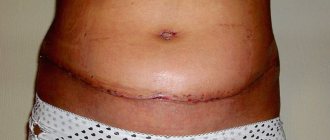
With this type of intervention, the deformation is minimal: the breast remains the same size, the decrease in volume is insignificant and almost imperceptible - after removal of the tumor, the breast returns to the original size that the patient had before the tumor developed.
The stitches are cosmetic in nature; over time they fade and become almost invisible.
The functionality of the glands is also preserved, so natural feeding is possible after childbirth.
Recovery period
- After sectoral resection of the mammary glands, the patient is discharged from the hospital on the third day. The drainage is removed within 2–3 days. The bandage remains on the body for up to 5–7 days - over the next week it is necessary to go to dressings, then it will be enough to treat the postoperative sutures with a green diamond solution
- It is necessary to conduct a preventive course of antibacterial therapy in order to prevent the development of purulent complications
- Then, to heal the sutures and prevent severe tissue scarring, collagen and silicone creams can be used as prescribed by the doctor, accelerating the regeneration of the ointment
- It is also recommended to enrich the diet with animal proteins, microelements and vitamins to overcome postoperative stress and replenish iron reserves lost in the blood
It is important to monitor your condition during the first week after the intervention:
- Measure the temperature 2 times a day (it can be raised to low-grade levels in the first 2 days after surgery - this is a physiological reaction)
- Observe the infection safety regime
- Monitor the condition of the skin around the wound and the intensity of pain
If the temperature persists for more than 3 days or if it increases significantly, pain appears, severe redness of the skin, swelling of the chest, copious or purulent discharge (opaque, white-yellow or greenish) from the wound, immediately contact a hospital or your doctor.
Advantages of carrying out the procedure at MEDSI:
- Reception of mammologists and gynecologists of international class
- Full hardware examination, cancer search
- Segmental resection of mammary glands by leading surgeons in Moscow
- Comfortable stay in the hospital hospital
- Assistance with accommodation of out-of-town patients during the rehabilitation period
- Feedback from specialists
It’s easy to sign up for a consultation – just call 8 (495) 7-800-500 (calls are accepted 24 hours a day).
Diagnosis of the accessory lobule of the mammary gland
Pathology can be detected and confirmed using available methods. Namely:
| Method | Diagnostic features |
| Inspection and palpation | Can give general ideas about education. But if the lobule is undeveloped, there are no changes in it, it is impossible to identify it. |
| Ultrasound examination of the mammary glands | Often, an additional lobe is discovered by chance, especially if the woman is not bothered by it. |
| CT or MRI | They are used less frequently, for example, to clarify the nature of the formation or suspicion of a tumor. |
| Mammography | To identify it, it is necessary to specifically examine the axillary region (or subclavian, etc.). |
| Puncture | If necessary, especially in case of mastopathy, a puncture can be performed followed by a histological examination of the obtained material. |
What is our hospital like?
24/7, comfortable, professional, caring. KSM provides high-quality medical care at all stages of treatment, including recovery during the rehabilitation period.
Each patient is provided with a comfortable room and basic necessities - towels, robe, slippers, etc.
Attention! Each room has an emergency call button for medical staff.
But most importantly, you can count on the maximum effectiveness of the surgical treatment performed in accordance with international standards, as well as full rehabilitation according to an individual program.
To make an appointment, call or submit a request online.
Is education dangerous for health?
The extra lobe itself is not dangerous to a woman’s health. Many are afraid that it will eventually turn into a malignant tumor. In fact, the probability for this pathology is exactly the same as for all other women.
The difficulties are as follows. In some situations, pathological processes in this area may be diagnosed late due to a somewhat atypical clinical picture. As a result, you can get an advanced process, for example, with inflammation - an abscess, with a tumor - stage 3 - 4, etc.
To avoid this, it is necessary to undergo regular examinations with a gynecologist and, if there is pathology of the mammary glands, with a mammologist. And if complaints arise, seek medical help immediately.
Treatment of diseases associated with enlarged axillary lymph nodes
Based on the diagnostic data, the doctor will prescribe treatment for the underlying disease that caused the enlarged lymph nodes:
- conservative treatment of mastopathy, mastitis;
- if necessary, surgical treatment (from opening a boil or abscess to serious operations on the mammary gland);
- chemotherapy if malignant tumors are detected;
- treatment of other identified diseases.
Early diagnosis of diseases that cause axillary lymphadenopathy, including breast pathology, provides the best results in treatment. We diagnose the cause of enlarged axillary lymph nodes with an accuracy of more than 90% thanks to a comprehensive expert examination, which includes ultrasound, mammography, and MRI.
Avoid prolonged enlargement of lymph nodes. This will lead to a decrease in the efficiency of the lymphatic system, which will be very difficult to restore. By making an appointment with a doctor, you can return your body’s functioning to normal and avoid the development of concomitant diseases.
Is it necessary to have surgery?
If the additional lobule of the mammary gland is not complicated by anything (abscess, cyst, tumor, etc.), there is no need to remove it. Even if it brings tolerable discomfort during breastfeeding or on the eve of menstruation, this is not an indication for surgery.
Removal of the additional lobe is recommended in the following situations:
- with large sizes;
- if it is accompanied by severe pain, swelling;
- if a cyst, fibroadenoma or even a malignant tumor is found in it;
- at the request of the woman, if it brings her psychological or cosmetic discomfort.
If a girl is bothered by some minimal symptoms, you can try treatment with drugs intended for the treatment of mastopathy. This way you can reduce the manifestation of pain, discomfort, swelling, and slightly reduce the size of the formation. Using properly selected hormonal contraception also helps relieve all symptoms.
Watch the video about benign breast tumors:
Removing the extra milk lobule
Operations to remove additional lobules of the mammary glands can proceed in different ways. It all depends on the size of the education, the desire of the woman, her age, whether she is going to give birth, etc. The approach is individual in each case.
Similar operations are performed by both oncologists and plastic surgeons. It is better to give preference to the latter if the lobule is not removed due to a tumor or abscess, since these doctors in most cases have more experience in correcting “extra” areas of skin, etc.
How is the operation performed?
With age, glandular tissue both in the mammary gland and in the accessory lobule is replaced by adipose tissue. This influences which surgery should be preferred. If, based on the results of the examination, it is determined that most of the lobule is adipose tissue, conventional liposuction can be performed. To do this you will need to make one small incision. Upon completion of the operation, it sometimes becomes necessary to install drainage to drain fluid for up to 3 to 5 days.
If the operation is performed at a young age, glandular tissue most likely predominates in the additional lobule. In this case, the entire gland should be removed. The steps of the operation are approximately as follows:
- Tissue incision over the accessory lobule or other cosmetic access.
- Removal of glandular structures and fatty tissue.
- If necessary, tissue plastic surgery, including removal of excess skin flap.
- Layer-by-layer suturing of tissues, if necessary, installation of drainages for the outflow of pathological fluid.
Recovery after deletion
The recovery period depends on the volume and type of surgery and ranges from a week to a month. For some time after the intervention, the woman may feel discomfort in this area. The main recommendations for preventing complications after surgery are as follows:
- It is necessary to follow all doctor's recommendations for treating the wound and taking medications. This will help prevent the wound from becoming infected.
- It is advisable not to become pregnant for 6 to 12 months, otherwise it may cause a relapse, especially if it was technically difficult to remove all the glandular tissue.