If after rhinoplasty surgery, bone grinding has formed on the bridge of the nose, then compensatory mechanisms did not help. This growth is the body’s desire to create a certain margin of safety after surgery. This is why excess bone fibers grow in the operated area. These accumulations are called “callus”. This phenomenon is quite rare, but every person who undergoes rhinoplasty is at risk.
According to statistics, approximately 12-15% of patients are susceptible to this complication, and 30% require reoperation. It is believed that callus after rhinoplasty is not life-threatening, but it should still be removed, as it can cause chronic pain and other unpleasant complications.
It takes about a year for such a defect to form. This is the result of the standard process of tissue regeneration after correction, if bone was affected during it. If the surgeon did not manipulate the bone frame, then a callus will not form.
The structure of this defect is connective tissue that grows at the junction of bones. And the growth process itself takes place in several stages:
- A provisional callus is formed;
- Osteoid tissue appears;
- Instead of bone fibers, connective tissue fibers appear.
In some cases, cartilage forms and from this cartilage forms bone. To prevent such a defect from appearing, after rhinoplasty you need to strictly follow the surgeon’s advice and recommendations.
Causes of calluses
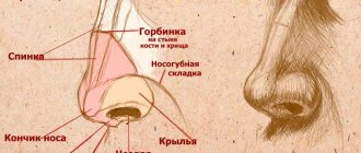
The framework of the nose consists of bone and cartilage. During the operation, certain elements are removed, and therefore such an operation is always associated with damage to the internal nasal structures. As a result, a reaction occurs in the form of activation of the body’s defense mechanisms. Its size depends on the degree of tissue injury and the individual characteristics of the body.
The main reasons for the appearance of formations:
- Individual reaction of the body to damage;
- Little experience and low qualifications of a plastic surgeon.
How is nasal septum surgery performed?
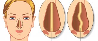
Today, plastic surgeons use 2 main methods of performing rhinoplasty for a hump nose: open and closed. With a closed technique, the incision passes through the nasal mucosa, with open rhinoplasty - through the columella. In most cases, when it is necessary to surgically correct a protrusion on the nasal septum, doctors use the open method, since thanks to this access the surgeon has complete control over the process and can create a nose that is ideal in shape and symmetry.
Hump rhinoplasty takes place in 4 stages:
- Septoplasty is a plastic surgery that allows surgical correction of the nasal septum. Thanks to septoplasty, the surgeon improves nasal breathing and relieves the patient of numerous problems: chronic runny nose, nasal congestion, snoring.
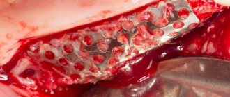
- Correction of the nasal bridge. During this stage of plastic surgery, the surgeon uses a scalpel and an osteostomy - a special surgical instrument that allows you to remove the protruding part of the bone.
- Stabilization of the nasal septum. The third stage of rhinoplasty involves fixing the bridge of the nose, expanding the nasal passages through the installation of grafts that the plastic surgeon creates from the tissues of the nasal septum. As a result of the operation, nasal breathing can be improved.
- Fixation of the tip of the nose. The final stage of septum rhinoplasty involves the installation of a graft in the area of the wings of the nose, thanks to which it is possible to stabilize the tip of the nose. The graft is sewn to the nasal septum, which gives the organ additional strength.
How long does facial swelling last?
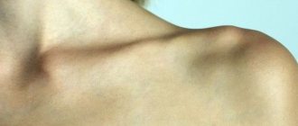
It is difficult to give exact dates, since, as mentioned above, there are a number of factors that influence healing. Conventionally, rehabilitation takes several stages. The first, during the first seven days after surgery, is the most unpleasant. Edema develops and reaches its peak by the third day after correction. During this period, there may be severe pain and slight bleeding, and there may be difficulty breathing through the nose. Swelling is expressed not only on the nose, but also in other areas of the face, in particular on the cheeks and chin.
By the third week, noticeable swelling of the face after rhinoplasty has already subsided, including those on the nose. Secondary edema remains. In most cases, it is invisible to the patient and others, but remains visible only to a professional, that is, a surgeon. 2-3 months after the operation, the swelling is no longer visible at all. But in general, the general recovery time is as follows: swelling goes away from the root of the nose in 2-3 months, from the back - in 4-6 months, from the tip - in a year and a half.
What is callus and how does it form after rhinoplasty?
Bone callus after nose correction is the growth of connective tissue at the site of a violation of the integrity of the bone after surgery. A growth forms on the bridge of the nose as a compensatory mechanism to increase bone strength. It does not pose any threat to life, but is an aesthetic defect.
Callus is not dangerous to health, but it can cause pain and various complications. Usually the bone tissue after surgery is restored within a year. During rhinoplasty, it can only appear if the bone is affected during surgery and its integrity is disrupted.
Stages of callus formation
There are three stages in the process of formation of a growth on the nasal bone:
- Formation of a provisional callus - at the site of a violation of integrity, a primary callus is formed from granulation tissue. This happens in the first 7-10 days after rhinoplasty. During this period, there is a divergence of bones at the fracture site and the transformation of granulation cells into connective cells.
- Bone tissue formation occurs 10-30 days after correction. During this period, connective tissue is transformed into bone cells - osteoblasts, between which there are collagen fibers. A bone callus is formed.
- Replacement of osteoid tissue with connective tissue fibers. Normally, after a month, the bone tissue begins to increase in the number of osteoblasts, after which at week 24 the bone is completely formed. However, sometimes the connective tissue continues to grow over time, forming a hyperplastic callus.
The likelihood of such a pathology is higher at a young age, when bone structures are not yet fully formed, and the growth zone and regeneration processes are extremely active. Therefore, rhinoplasty is recommended after the final formation of bone and cartilage tissue, not earlier than 25 years.
Types of calluses
The formation of callus depends on many factors, which include physical health, age, extent of surgical intervention and individual characteristics of the body. There are three main types of bone callus formation in rhinoplasty:
- Periosteal - formed from the outside, occurs at the point of fusion of the nasal bones. Does not apply to pathological processes. During formation, osteoblasts—bone cells—are actively formed.
- Intermedial - an intermediate callus that grows between the outer and inner parts of the fracture. This callus connects scattered bone fragments and restores its integrity. If the plaster cast after rhinoplasty is applied correctly, fits tightly to the bone, and does not allow bone fragments to move, then callus formation proceeds correctly. If there is a displacement, then there is a possibility of build-up formation.
- Endosteal is an internal bone callus formed from the endosteum. There are no blood vessels on this part of the bone, so the callus gradually grows and bulges.
The first to form are external and internal calluses. They temporarily connect the fragments and create conditions for subsequent bone fusion. They resolve gradually, being replaced by the resulting periosteum and bone tissue. The intermediary callus forms later and firmly connects the parts of the bone. This is a normal process. When the regeneration process is disrupted, the connective tissue grows excessively and extends beyond the bone fragments.
Causes
Overgrowth of connective and bone tissue occurs as a result of improper fusion of the nasal bones.
The cause may be improper repositioning of bone fragments during surgery, but most often it is the result of mechanical trauma during the regeneration period, which led to bone displacement or damage to the soft tissue of the nose around the operated organs, for example, the vessels feeding the bone. To fix the nasal bone fragments, surgeons apply rhinological splints and splints (plaster casts). The more securely the bones are fixed, the more successful the regeneration. During the recovery period, this part of the face is very vulnerable, and the slightest mechanical impact can lead to disruption of recovery processes.
The formation of bone tissue is also influenced by the state of the body itself and its various systems. The recovery processes are influenced by hormonal status, the state of the immune and vascular systems, the activity of metabolic processes, as well as the tendency to form keloid tissue.
Diagnostics
Diagnosis of callus is based on examination and radiography. On an x-ray, the growth appears as a membrane surrounding the area of bone damage. The image helps determine the exact location of the growth, its size, and develop treatment tactics.
Prevention during the rehabilitation period. Recommendations from a plastic surgeon
To prevent excessive growth of connective tissue and the formation of callus in the surgical area, a number of rules must be followed during the period of restoration of bone integrity. The main one is to avoid injuries, take measures to reduce the inflammatory process in the correction area and ensure improved tissue trophism.
During the recovery period, which lasts three months, you must be under the supervision of a surgeon. The doctor monitors the formation of bone tissue. This is the most important follow-up stage after rhinoplasty. Final formation occurs after 6-9 months, and ends after a year, when the connective tissue resolves and bone and cartilage tissue is restored.
To minimize the risk of calluses, I recommend following simple rules:
- sleep only on your back for the first three months;
- do not wear glasses, including light glasses with tinted lenses;
- lifting and moving heavy objects is also prohibited for the first three months;
- do not blow your nose for two weeks;
- exclude heavy physical activity in the first six months;
- sneeze with your mouth open;
- monitor the correct position of the splint, its tightness, do not move or remove it yourself;
- start playing sports after the surgeon’s permission;
- at the first symptoms of hyperplasia, consult a doctor;
- Avoid infections and inflammatory diseases in every possible way, especially rhinitis and sinusitis;
- for the first two months do not visit the solarium, do not sunbathe;
- during the same period, exercise in the pool is contraindicated;
- exclude cold showers, saunas, hot baths.
I do not recommend air travel for the first six months. During the year, you need to avoid conflict situations, stress, and other psychological trauma.
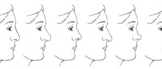
Causes of formation and methods of removing a nasal hump
The reasons for the appearance of a cosmetic defect on the nose are as follows:
- Heredity. A hump of the nose is a characteristic feature of the facial features of representatives of some nationalities. In this case, many people perceive education as a highlight of appearance. It gives a noble appearance to the face;
- Traumatic injuries to the bones and cartilage of the nose, after which they do not heal properly. As a result, the appearance of the nose deteriorates;
- Consequences of incorrectly performed previously plastic surgery.
Plastic surgeons at our clinic use modern techniques to remove the hump on the nose. The price of the operation depends on the complexity. Doctors work simultaneously with bone and cartilage tissue. They use the following techniques: contouring and rhinoplasty.
You can remove a small hump by performing contour plastic surgery. The aesthetic surgeon injects a special substance under the skin of the bridge of the nose. It evenly fills the space and aligns the line of the nose. The effect lasts for one or two years. After this, repeated plastic surgery of the nasal hump is required. As a result of surgical intervention, the aesthetic appearance of faces improves. If a patient has problems with nasal breathing, he is offered rhinoplasty. The nasal hump is eliminated simultaneously with the correction of the nasal septum.
Rhinoplasty of the nasal hump (the price is higher than the previous procedure) is a surgical operation during which the surgeon excises the nasal bone and cartilage tissue with special instruments. It solves the following problems:
- Aesthetic – improves the appearance of the nose;
- Physiological – restores nasal breathing;
- Medical – relieves the patient from snoring and nasal congestion.
How to remove callus after rhinoplasty?
When bone tissue grows on the nose, it appears to be of several types at once. Manifestations of this defect include:
- The appearance of a hump;
- Subsequent deformation of the noma, and the change can be clearly visible from the front;
- Swelling.
Naturally, such manifestations of beauty do not add to the beauty and dissatisfaction with the results of the operation appears. These phenomena worsen the aesthetics of facial features. If the defect is severe, repeat rhinoplasty will have to be performed to remove bone tissue at the site of the growth.
The appearance of the first symptoms makes patients very nervous and consult a doctor. At the same time, there is a fear of revision rhinoplasty. Please note that this is done very rarely. First, the patient should try preventive measures that help cope with the problem for most people operated on in this area.
By the way, the doctor can recommend some measures even before the operation. Therefore, carefully observe his wishes and instructions. But the main measures, of course, come precisely during the rehabilitation period. During this time, you must visit the surgeon at least five times so that he monitors the regeneration process and can promptly notice certain changes in the process of tissue healing.
Secondary correction is prescribed, as mentioned above, by approximately 30% of the total number of people with complications of this type. First, the doctor must review the picture in detail and study the features of the process. During the operation, special measures are taken by the doctor, taking into account the characteristics of the human body. This helps prevent secondary growth of bone tissue.
What do we have to do?
At the first signs of callus after rhinoplasty, certain measures are mandatory. First of all you must:
- Visit a doctor. In general, even under normal circumstances, visits are required in the first, second, third, sixth and twelfth months after surgery. In case of force majeure, an additional visit can be made. If a deviation is detected, the doctor will prescribe treatment.
- If the doctor has prescribed treatment, then you must follow it strictly and strictly according to schedule. Otherwise, the therapy may simply not give the desired result and there is no point in blaming the doctor’s abilities in such cases.
- People under 18 years of age are susceptible to tissue hypergrowth. Therefore, they are not recommended to undergo surgery. This is explained by the fact that the body continues to develop, and with it the tissues. With this type of intervention, by removing one defect, there is a risk of getting much larger problems. The patient's readiness for surgery is determined only by the plastic surgeon. An experienced person will refuse if the conditions for the operation do not meet. In such cases, we do not recommend looking for someone else who will allow it.