The desire for ideals was born almost simultaneously with the appearance of the first man on Earth. To achieve bodily perfection in different eras, people sometimes used unexpected substances, trying to give additional volume to those parts of the body that lacked it. However, these experiments were dangerous to health and did not lead to the desired result. In the second half of the twentieth century, the idea of a safe and effective increase in volume finally found its embodiment in filleting.
NATALIA MIKHAILOVA , Ph.D., dermatovenerologist, cosmetologist, associate professor of the department of skin diseases and cosmetology of the Federal Faculty of Postgraduate Education, Russian National Research Medical University named after. N.I. Pirogova, scientific director of the International Academy of Aesthetic Medicine Natalia Mikhailova, Russia.
LYUDMILA SOBOLEVA , dermatovenerologist, cosmetologist, scientific editor of the publishing house "MEDESTHETIC-PRESS", member of the expert council of Suisselle (Switzerland), Russia.
The material was provided by the Natalia Mikhailova Academy with the support of the National Society of Mesotherapy (NOM) and the Eurasian Society of Injection Techniques Specialists (ESOSIM).
Filler degradation
The past century was marked by the highest discoveries and achievements in the field of chemistry, pharmacology, and medicine. The most dynamic process of creation and clinical use of injection implants occurred in the twentieth century. Over this century, a path has passed from permanent implants through semi-permanent to biodegradable ones.
Permanent implants (silicone, polyacrylamide gel) were, of course, a breakthrough compared to previously used liquid paraffin injections. But experience has shown that the duration of the effect can easily turn into disaster: the occurrence of complications that are difficult to treat. Non-degradable implants are in some cases limited by a dense fibrous capsule. It may be noticeable or even noticeable, but in principle this is a favorable scenario. However, this does not always happen and not with everyone. Much more dangerous are materials that are poorly contained in a living organism. They can literally “saturate” the tissue, so removing such an injectable implant is a complex, painstaking work, a traumatic operation that absolutely does not guarantee the complete elimination of the drug. As a result, tissue deformations, severe scarring and other problems with surgical correction are quite likely.
Delayed complications
A feature of non-degradable drugs is late complications. for example, 5 or 10 years after the injection. This can happen against the background of a general illness, changes in living conditions, or climate. It is not uncommon for patients to seek re-correction after injection of non-degradable fillers. Exposure to radio frequencies or deep heating, intense massage, injections - all this can provoke the appearance of late infectious complications in the area of insertion of a non-degradable implant.
Despite the active dissemination of information, the number of patients injected with biopolymers continues to grow. Unfortunately, this trend has gained momentum again over the past couple of years, which can be associated with the economic crisis and a natural increase in prices for biodegradable implants. At the same time, advertising of biopolymers uses such tricks as “modern silicone”, “absorbable polymer” - alas, these names most often hide those same non-absorbable fillers. The injection itself is cheaper and the effect lasts longer. The only risk that nothing is said about is the cost of one’s own health and appearance.
Anatomical structure of the face
The key to understanding the causes of various complications after filler injection is a good knowledge of the structure of the facial vascular system. In general terms, the anatomical structure of the human face can be divided into 5 layers:
- Skin (epidermis and dermis)
- Subcutaneous fat deposits (Fig. 1)
- Facial muscles and SMAS (Fig. 2)
- Blood vessel system (Fig. 3)
- Scull
|
|
|
You should also take into account all procedures and surgeries that the patient has undergone previously, since they could affect the anatomical structure of the face and be a factor in the increased risk of undesirable consequences of the injection of fillers.
Collagen: safety test
The properties of bovine collagen and its relationship with the human body were studied for 20 years, after which the drugs received FDA approval and began to be used to restore soft tissue volumes. For a long time they were the most popular filleting product. Among the collagen fillers, there were preparations based on xenogeneic (bovine, porcine), homologous (human) and autologous (own) collagen. xenogeneic implants should be considered more dangerous - there is a risk of an allergic reaction, a sensitivity test is required. Collagen itself is a completely biodegradable substance. This is a favorable sign from a safety point of view, but the same circumstance requires frequent repeated injections. For this reason, collagen began to be combined with other substances - for example, polymethyl methacrylate granules. Thus, collagen-based drugs have acquired different biodegradation periods (from 1 year to infinity), but their safety profile has also changed: in addition to the potential risk of allergies, the likelihood of all complications of non-degradable drugs has been added. The direction of creating an autologous culture of fibroblasts with collagen and elastin fibers was considered quite promising: a fragment of the patient’s own skin was cultured. However, this labor-intensive method remained at the stage of clinical trials; new drugs began to combine high-quality production, ease of use and time-appropriate correction.
An exception
The category of semi-permanent fillers is of undeniable interest, often combined with concern. These include substances such as L-polylactic acid and calcium hydroxyapatite. A feature of this category is the stimulating effect, that is, in essence, the drugs cause noticeable fibrosis in the injection area. This property is used in cases where lifting of sagging skin and long-term replenishment of volume deficit are necessary. One of the priority indications for the use of drugs in this group, in particular L-polylactic acid, is facial lipoatrophy in HIV-infected patients (on the one hand, they suffer from severe soft tissue deficiency, and on the other hand, they do not demonstrate a strong immune response to administration drugs). These drugs already have quite a long history, and it has both obviously positive connotations and a share of negative reviews. As a rule, analysis of unsuccessful experience situations shows frequent violations of injection techniques and patient selection rules. The difficulty of working with these drugs is due to their mechanism of action. Of course, they provide a certain volume effect, but the stimulating effect on the dermis/hypodermis with the formation of fibrosis is still more significant. Drugs in this group are most often diluted before use. The trend of recent years persistently shows us the use of less and less concentrated solutions. A more liquid solution undoubtedly has a less pronounced effect. But at the same time, it clearly stimulates fibrosis without the risk of overcorrection.
Soft fabrics: reinforce without waiting for sagging
Hyaluronan is the name given to the salt of hyaluronic acid, and in preparations it is found in the form of sodium salt. The history of drugs based on hyaluronic acid (HA) began long before the creation of fillers. This substance was first isolated from biological material almost a hundred years ago [4], and the description and the term itself belong to Carl Meyer and John Palmer. In medicine, hyaluronan began to be used as a wound-healing agent, and then the scope of use of HA-based drugs began to rapidly expand. In aesthetic medicine, hyaluronan initially appeared as an external agent: in the form of serums, masks, and cream components. The use of HA in cosmetics is associated with the main feature of the molecule: it has extremely high hygroscopicity and is capable of retaining a significant amount of water. In tissues, this process is reversible; the destruction of the molecule leads to the release of a significant amount of moisture, which has a moisturizing effect on the skin.
In the 80s last century, the first injectable drugs based on HA began to appear. Today, there are a great variety of injectable drugs based on HA. Some of them are very similar, others are significantly different. Of course, the primary quality factor is a reliable source of hyaluronic acid raw materials. Considering the volumes of HA used today in a wide variety of branches of medicine, bacterial synthesis is used as a source of hyaluronan. The overwhelming majority is synthesized by non-pathogenic strains of streptococcus [4], and HA produced by Bacillus Subtilis is also found. HA can have different molecular weights (length, or rather volume of the molecule), and polymers of different sizes can have different, sometimes opposite, effects.
Among injectable preparations based on hyaluronic acid, two categories can be distinguished: therapeutic group and soft tissue fillers - fillers. The first group includes mesotherapeutic options, biorevitalizants (sometimes they are also called skinboosters), bioreparants. These drugs exist in living tissues for a fairly short period of time, since within a few days after injection they are broken down into monomers by the enzyme hyaluronidase. Injection of these drugs allows you to obtain the effect of hydration of the dermis, improving trophism of the skin in the injection area, increasing turgor and smoothing wrinkles. Such drugs are usually used in course protocols.
The second group is fillers, the biodegradation period of which is calculated in months. Filler injections are carried out to fill wrinkles and folds, restore volume in areas of deficiency, and tighten - “reinforce” the skin.
Volume and shape
Historically, the process of creating drugs began with biphasic fillers. As a transport medium, stabilized HA was immersed in the native solution to ensure fluidity and create the optimal consistency of the drug. The significant volume of the transport medium determined the peculiarities of working with the drug: the occurrence of pronounced edema at the implantation site, followed by a significant loss of volume and the need for additional correction. Over time, the technology for creating biphasic fillers has changed in such a way that it has been possible to reduce post-procedure swelling and subsequent volume loss. This significantly increased the comfort of using the drugs. Biphasic fillers are characterized by the ability to retain volume and shape well for a long time. For this reason, they are very appropriate in procedures for recreating/restoring volume.
The range of biphasic fillers is always represented by preparations containing the same concentration of HA - most often 20 mg/ml. The difference in the properties of the drugs is achieved due to the particle size. Smaller ones (accordingly, there will be more of them per unit volume) will degrade faster; these drugs are administered more superficially and correct relatively small defects. Larger particles are present in denser preparations; they degrade longer, meaning a longer clinical effect is achieved. These drugs are injected into deeper layers: subcutaneously or into the deep fatty layer, onto the periosteum. Biphasic drugs can provide not only volume restoration, but also pronounced lifting.
Location of blood vessels in dangerous areas
The most dangerous complication associated with dermal filler injections is venous or arterial occlusion, which can lead to ischemia and subsequent skin necrosis and/or vision loss. All these phenomena are extremely undesirable and require immediate medical intervention. Arterial occlusion can lead to the formation of ulcers and scars, and occlusion of the ophthalmic artery can lead to loss of vision.
The causes of arterial or venous occlusion can be the injection of filler directly into the vein/artery, damage to the blood vessel, or external pressure exerted on it by the injected filler or the formation of edema. In general, blood vessel occlusion and necrosis are considered rare complications.
Rice. 4. Danger zones when injecting fillers
Figure 4 shows the main danger zones of the face, which should be avoided when injecting fillers. In the cheek area it is:
- facial artery;
- transverse artery of the face;
- buccal branch of the maxillary artery;
- infraorbital artery;
- zygomatic branch of the buccal artery.
In the area of the glabella, due to the small diameter of the vessels and insufficient collateral circulation, the injection of fillers should be done with extreme caution. Here the areas of increased danger are:
- frontal artery;
- supraorbital artery.
Dangerous areas in the nose area include:
- external nasal artery;
- angular artery.
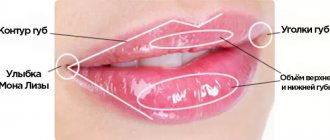
In the perioral zone it is:
- superior labial artery;
- inferior labial artery.
In the temple area, fillers are injected with caution, avoiding the superficial temporal artery, its posterior parietal and anterior frontal branches.
Persistent molecule
Initially, HA was made more stable in tissues by combining it with DVS (divinyl sulfone). From 1993 to the present day, fillers have generally been stabilized by combining them with a chemical called epoxide, abbreviated BDDE (1,4-butanediol diglycidyl ether). BDDE forms bonds between chains of hyaluronic acid molecules, which changes their spatial structure and makes it unrecognizable to hyaluronidase. This allows the substance to be present in tissues much longer. The nuances of the stabilization (reticulation) process can be regulated by the technological process, which makes it possible to create a large assortment of unique drugs with different biodegradation periods.
Stabilized HA is subjected to a grinding process by homogenization or sieving. This stage determines one of the main properties of the filler - whether it belongs to the class of monophasic (homogenized) or biphasic (sifted) fillers.
Not painful and safe
A number of modern fillers are represented not only by stabilized hyaluronic acid, some contain additional ingredients on which the properties of the drug may depend. One of the most common is lidocaine. This well-known anesthesia drug is in some cases added to the noe. Of course, this does not affect the injection procedure itself in any way. But in the injection technique for such drugs, they usually recommend not the traditional retrograde, but a combination: first, a small amount of filler with an anesthetic is injected antegradely, and then the rest of the volume is injected retrogradely. From the point of view of tolerability of the procedure, this may be more comfortable, but it should be remembered that lidocaine is quite often an allergen and the patient’s medical history should be carefully ascertained. The second point is that lidocaine can cause tissue swelling and it can be difficult to evaluate the result immediately after the procedure. However, the topic of connecting with anesthetics is now quite popular; in some cases, specialists independently mix filler with lidocaine using connectors.
What small pieces
When creating monophasic fillers, the stabilized hyaluronic acid gel is crushed (homogenized). In this process, the gel consists of many particles of different sizes. Monophasic fillers within the same line may differ from each other in concentration or degree of modification, calculated as a percentage (hence, the number of cross-links per certain number of links in the HA chain). In general, the structure of monophasic fillers ensures the comfort of the drugs during administration and integration into tissues. The lifting effect and ability to retain volume are somewhat less pronounced than in biphasic gels.
allows you to reduce the severity of the inflammatory reaction in response to injection of the drug. Accordingly, this can provide the patient with a more comfortable post-procedure period. But it should be understood that the stimulating effect on the dermis during injection will be much less pronounced.
Possible complications: symptoms and consequences management
A change in skin color immediately after filler injection indicates vascular ischemia. Symptoms of arterial or venous occlusion, which can lead to skin necrosis, include discoloration or darkening of the skin (to a blue-gray hue), ecchymosis, reticular erythema, and severe pain at the injection site.
In cases of vascular occlusion or ensuing necrosis, the main goal is to stimulate blood flow in the affected area. The following methods can be used for this:
- immediate application of a warm compress,
- massage the affected area to dilate blood vessels and disperse the injected drug,
- taking aspirin,
- the use of external drugs that stimulate vasodilation,
- injection of hyaluronidase (in case of injection of hyaluronic acid fillers),
- injection of corticosteroids (anti-inflammatory/immunomodulatory),
- taking antibiotics,
- taking antiviral drugs (in case of threat of necrosis in the perioral area).
It is also possible to use hyperbaric oxygen therapy and laser procedures 3 months after the injection of the filler.
Cases of retinal artery occlusion are extremely rare and very dangerous. Symptoms of possible vision loss are sharp pain in the area of the affected eye immediately after filler injection, blurred vision, ptosis, headache, dizziness, nausea, and ophthalmoplegia.
Usually, in the case of retinal artery occlusion, vision loss is irreversible. The best way to prevent this extremely undesirable phenomenon is to avoid dangerous areas when injecting fillers, especially in the area of the glabella, forehead and upper part of the nasolabial folds.
Fillers of the future
By analogy with meso-cocktails, which are prepared ex tempore, the question arises: how evenly distributed are the additional components in the filler? Will it happen that at some stages of the procedure the tissue will receive a filler with an additive, and at others - an additive with a filler? Indeed, when preparing a cocktail with hyaluronic acid, this fact is obvious: at the beginning of the procedure, a more liquid drug is supplied, and at the end - a more viscous one. As a rule, cosmetologists have to take this nuance into account when choosing a specific protocol, the sequence of techniques, and repeatedly returning the needle to the same areas. Recently, this aspect has received considerable attention. And if in the creation of therapeutic drugs this issue has already been resolved with the help of compositions based on HA with components sewn onto it, then for fillers this will be a new page in history. It is likely that the traditional HA filler will soon be replaced by new combinations that combine the filling effect with a uniform and long-lasting therapeutic effect. It is possible that these combinations will include antioxidants - in this case we can get an ideal filler for skin with photoaging, stimulating factors - for example, polylactic acid, to achieve long-term restoration of soft tissue volume, peptides - so that, along with filling, they have a pronounced therapeutic action.
Principles for safe injection of fillers in hazardous areas
In rare, but still possible, cases of vascular occlusion and subsequent necrosis and/or loss of vision after injection of fillers, emergency medical measures are essential. Sudden sharp pain and/or skin discoloration are warning signs of vascular occlusion, ischemia, and impending necrosis. To reduce the likelihood of undesirable consequences of this nature, it is recommended to follow the following rules:
- inject filler in small doses,
- do not use anesthesia (including drugs containing epinephrine) near vascular bundles, so as not to cause vascular spasm,
- avoid the use of epinephrine to be able to quickly identify the cause of skin discoloration,
- use a small needle so that the filler enters the tissue in minimal doses,
- use biodegradable fillers (for example, hyaluronidase stimulates the rapid breakdown of hyaluronic acid fillers),
- when injecting filler, use the finger of your non-dominant hand to block the adjacent blood vessel,
- inject filler into the upper and middle layers,
- assess the level of pain of the procedure,
- monitor possible changes in skin color,
- If non-biodegradable fillers are used, administer them in small doses and take into account their high viscosity.
To prevent such a complication as vascular occlusion, I propose to use a simple and effective technique - an aspiration test: after inserting the needle into the correction zone and before starting to deliver the drug, it is necessary to perform a slight reverse movement of the syringe piston. If there is no blood, you can start administering the drug. This test allows you to exclude the drug from entering the vascular bed.