Forms of neuralgia of the facial nerve
- neuritis – minor inflammation of the facial nerve (or part of it),
- paresis is a partial lesion in which weakness of the facial muscles and slight numbness are observed. This disease usually falls into the neurological category. But, like complete paralysis, it can be a consequence of advanced neuritis,
- paralysis - complete defeat. This is a severe stage, in which there is numbness of the face (usually one part of it) and blocking of muscle motor activity.
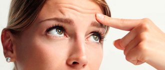
Symptoms of neuralgia
- tingling or sharp “sliding” pain,
- numbness of a certain part or the entire half of the face, lack of sensitivity and motor activity,
- drooping eyelids and skin around the eyes,
- drooping corners of the lips (on the side where muscle paralysis is observed),
- smoothing out wrinkles on the cheeks and forehead,
- pronounced facial asymmetry.
Causes of neuralgia
- viral infection or inflammatory processes (ear, throat, nose),
- hypothermia,
- inflammatory processes in the roots of teeth and bone tissue of the jaw,
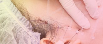
- nerve injury during tooth extraction or installation of implants,
- injury from a needle during the administration of anesthesia,
- jaw injuries,
- old age and natural aging of the body.
The facial nerve belongs to the group of cranial nerves (along with the trigeminal nerve, whose branches are the mandibular and maxillary nerves). It also consists of many branches that run through almost the entire human skull. Depending on which branch is affected, the numbness of a certain part of the face will depend. In relation to implantation or complex surgical interventions, it is the trigeminal nerve that can be damaged.
Exercises for paresis
Gymnastics for facial muscles in combination with ongoing therapy allows you to quickly cope with the disease. You can do the following exercises yourself in front of the mirror:
- upward movement of the eyebrows;
- frown;
- squint and open your eyes;
- smile without opening your mouth;
- smile with your mouth open;
- squint;
- lowering your head down, inhale and exhale, vibrating your lips;
- whistle;
- widen the nostrils;
- raise your upper lip, then lower your lower lip, exposing your teeth;
- put out a burning match;
- rinse your mouth with water;
- puff out one's cheeks;
- move air from one half of the mouth to the other;
- lower the corners of the mouth downwards with the mouth closed;
- stick out tongue;
- move around with your tongue sticking out of your mouth;
- protrusion of lips forward.
Remember: if symptoms of facial nerve paresis appear, be sure to contact a neurologist! Don't spread the disease, stay healthy!
Isaeva Rumiya Musaevna, neurologist at the “Healer” medical clinic in Khasavyurt.
Nerve damage after tooth extraction
As a rule, this problem arises during complex tooth extraction, namely, dystopic or impacted, that is, located inside the bone tissue and has not completely erupted. Removing such teeth requires sawing the bone and using special forceps. If the load is too heavy and the nerve is located close, the doctor may touch it.
The prognosis in such a situation is usually positive - the condition of the nerve is restored after therapy.
Diagnostics
Despite the fact that dystonic disorders, which include Bruegel's syndrome, are among the most common disorders of the motor system, the diagnosis of dystonia is not made very often. This is due to the fact that there are no clear diagnostic criteria for dystonia. In 2006, American neurologists published a number of characteristics that allow making a correct diagnosis and preventing underdiagnosis [4].
- Contractions of the same muscle group are repeated, and the speed of contraction remains constant.
- Hyperkinesis consistently affects one or more parts of the body. As the disease progresses, new parts of the body are involved or new movements appear.
- Changing the patient's posture can either increase or decrease hyperkinesis.
- Stress and fatigue increase the manifestations of dystonia, and rest and sleep reduce them.
- Against the background of hyperkinesis, the patient develops corrective gestures, with the help of which the patient tries to control dystonic phenomena.
Also in 2006, a classification of dystonias was published in the collection of official recommendations of the European Federation of Neurological Societies (EFNS), the use of which helps specialists correctly diagnose the disease, distinguishing it from other neurological movement disorders, such as tremor or chorea .
Trigeminal neuralgia as a complication of implantation
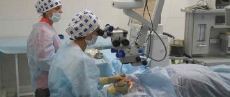
Unfortunately, one of the fairly common causes of damage to the facial (more precisely, trigeminal) nerve is dental implantation. According to recent studies, the frequency of such complications at this time does not exceed 2.95% (that is, it occurs in 5 out of 169 patients), and only 1.7% of patients are diagnosed with permanent neuropathy, requiring microsurgical correction of the situation1. If the implant is incorrectly selected in length, with increased pressure during its installation, there is a high risk of damage to one of the branches of the trigeminal nerve (passing on the lower or upper jaw). The result is pain or numbness in a certain area, long-term rehabilitation.
Injuries are caused by compression of a given nerve, its stretching, partial or complete damage. In the case of sprain or compression without compromising the integrity of the trigeminal nerve, the sensations pass quite quickly - within up to 7 days as blood circulation in the bone tissue improves. But if there was partial damage to the fibers of the nerve bundle, then the rehabilitation process can take from 14 days to 6 months. In cases of severe trauma, implants may need to be reinstalled because the body of the implant interferes with the restoration of axons (processes) in the nerve bundles.
In our clinic, before the operation, careful computer planning of the placement of implants is carried out, after which surgical templates are printed, protecting the patient and the doctor from incorrect installation of implants. Immediately, or the next day after the operation, a control computed tomography is performed, at which the control commission (consilium of doctors), in accordance with international quality assessment protocols, compares the placement of implants with the original plan and makes a conclusion about the success of the operation, or makes recommendations to the attending physician on the need to take additional measures.
Important! Do not confuse nerve damage with compression on it that occurs in the bone as a result of swelling after the installation of implants located close to the nerve endings. Compression goes away along with swelling and numbness in this situation is a normal reaction. Most often, such symptoms typically appear 2-3 days after multiple implantations; they are a normal reaction to tissue injury when screwing in implants and damage to intraosseous capillaries. Improvements will be noticeable almost immediately after installing the prosthesis on the implants and activating the chewing load, and with it the restoration and blood circulation in the bones.
Please note that there is no point in hiding the fact of damage to the trigeminal nerve for the doctor and the clinic; it will reveal itself anyway - this is the first thing. Secondly, even if this happened, the consequences are eliminated by properly selected therapy and joint work between the doctor and the patient. Yes, if damage occurs, then the patient will have to undergo quite a long period of rehabilitation (up to 6 months), but in difficult clinical situations of complete absence of teeth and low quality bone tissue, the doctor works in very limited conditions, so, unfortunately, some consequences but not life-threatening and health dysfunction cannot always be avoided.
Expert opinion
Nikolay Vladimirovich Namdakov Maxillofacial surgeon, implantologist, orthopedist Work experience 17+
“During one-stage implantation, we very often use elongated implant models that are installed in the deep parts of the bone tissue. Naturally, this increases the risk of damage to the nerves, which in this case are located very close to the implants. However, in our work we use three-dimensional visualization - we carefully study the implantation process in a computer program, where we set all the parameters of the jaw system of a particular patient. This allows us to eliminate the possibility of damage to the facial nerve during surgery, even in difficult clinical conditions of bone atrophy.”
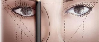
Correction of facial asymmetry
If flaws in the form of asymmetry suddenly appear in your face, this is not yet a reason to radically change your appearance. You can just use cosmetics and place the accents correctly. In this case, you need to use foundation and correctors. They can be oily or dry. In addition, she uses highlighter in her work. It is needed to visually hide wrinkles or highlight the necessary part.
The asymmetry of the eyes will not be so noticeable if they are not outlined with bright eyeliner. It is better to have tones close in color on hand, to make a soft transition from one color to another. Pencils with a contrasting color will also come in handy. On the inside of the eye, which we want to visually enlarge, it would be good to apply a lighter tone. On the eyelid of the second eye we apply an almost invisible line of dark color.
If the eyes, in your opinion, are still asymmetrical, then you need to place emphasis on the eyelashes and eyebrows. Eyebrows need to be given the perfect shape and arch. Latisse can enhance your eyelashes. A brown pencil can always highlight light eyebrows. And if you pluck an eyebrow that is higher than the other and draw on the eyebrow line with a pencil, your face will take on a completely different expression.
You need to have foundation creams on hand to be able to visually change the shape of your nose. The golden rule: a dark tone is applied to those places that need to be hidden or visually reduced. A lighter tone is used to highlight certain parts of the nose. To make the nose look good, you need to draw a line along the nose in a tone that is darker than natural. And leave a light light highlight on the wings of the nose and its tip.
A contour pencil helps give the lips the desired shape. And then the lips visually lose their asymmetry. You can correct your cheekbones with regular blush. To do this, you need two half-tone blushes that are close in color. They are applied along the cheekbone line, and their heights are different.
Treatment of lesions of the facial or trigeminal nerve
The first thing to do (if the cause of the lesion is not known) is to undergo a full diagnosis and find out why this problem occurred. This will allow the doctor to choose the right treatment tactics.
It is very important to consult a doctor as soon as possible if you experience the first symptoms of complications after tooth extraction or implant installation. If treatment is started promptly, complete restoration of muscle functionality can be achieved. It is also important that if numbness persists for 3 months and no measures have been taken, it will most likely not be possible to restore the affected nerve, since persistent degenerative changes develop in it.
Diagnostics
- a blood test to check for a viral or bacterial infection that could cause nerve damage,
- CT or MRI of the skull and brain to determine the affected area,
- electromyography, which involves direct impact on nerve endings to determine the level of disturbance in the passage of impulses along the nerve.
Treatment
- drug therapy: this group includes anti-inflammatory, painkillers and anticonvulsants, as well as antidepressants and sedatives.
- physiotherapy: facial gymnastics, massage performed by specialists, electrical stimulation and acupuncture,
- microsurgery: surgery is performed to restore the integrity of the damaged nerve. This is a fairly serious surgical procedure that should be performed by a professional surgeon. Surgery is an extreme method of solving the problem; it is needed only in case of serious damage and if the first two options did not bring any result.
Clinic and course
Bruegel syndrome first appears at the age of 50–60 years. Women suffer from this syndrome three times more often than men. Typically, the onset of the disease is accompanied by hyperkinesis of the orbicularis oculi muscle (blepharospasm) followed by oromandibular dystonia - hyperkinesis of the muscles of the face, jaws, pharynx and tongue.
The etiology of Bruegel's syndrome is currently unknown. The syndrome rarely occurs in children, affecting mainly older people. It is possible that the appearance of dystonia is associated with an imbalance of the main neurotransmitters in the brain and disturbances in the signal transmission system. Most likely, this imbalance is genetically determined.
Facial hyperkinesis manifests itself spontaneously - first, frequent blinking occurs, and gradually the closure of the eyelids becomes longer and longer. Blepharospasm is usually symmetrical - it affects both eyes at once, but there are cases when only one half of the face is affected. Blepharospasm is accompanied by redness of the face, increased breathing (dyspnea), and corrective gestures that occur when the patient tries to fight the symptom that has arisen. Some patients experience constant lacrimation, while others, on the contrary, suffer from increased dry eyes.
There are a number of paradoxical kinesias - motor activities that lead to a decrease in the manifestations of symptoms of facial paraspasm. These include sucking on lollipops and smoking. Alcohol intake, darkness, and closing one or both eyes also have a suppressive effect.
Usually, patients are able to find a position of the eyes in which they feel relief - the symptoms of blepharospasm practically disappear with half-closed eyelids or moving the eyeballs to extreme positions.
Many patients suffering from Bruegel (Meige) syndrome become unable to care for themselves due to the resulting “functional blindness.” The resulting hyperkinesis of the orbicularis oculi muscles leads to the inability to see normally, despite the fact that the vision in such patients is not impaired.
Oromandibular dystonia - dystonia of the mouth muscles - can appear several years after the syndrome manifests itself. This period can be up to 20 years, and some patients do not live to develop the generalized form of Bruegel's syndrome.
With oromandibular dystonia, the muscles of the lower jaw, cheeks, tongue, and pharynx are affected. In rare cases, spasms affect the respiratory and neck muscles. Hyperkinesis of these muscles leads to the appearance of spontaneous grimaces, involuntary movements of the lower jaw - opening and closing the mouth, protruding the tongue, torticollis, which is a consequence of spasm of the cervical muscles.
The progression of the disease leads to the development of speech disorders - from voice changes to complete dysarthria. Eating is also difficult. In most cases, with Bruegel's syndrome, oromandibular dystonia occurs together with blepharospasm. However, as mentioned above, these facial hyperkinesis can be symptoms of other diseases in the field of neurology (for example, essential tremor), which significantly complicates diagnosis. The diagnosis must be made taking into account all the symptoms of the disease.
The so-called “lower Bruegel syndrome” is much less common. In this case, patients develop dystonia of the muscles of the lower part of the face, and blepharospasm does not occur at all.
In 30–80% of cases, patients suffering from Bruegel's syndrome develop dystonia affecting other parts of the body. The most common writer's cramp is tonic tension of the muscles of the upper extremities, dystonia of the pharynx and larynx.
It is important that the emergence and progression of Bruegel syndrome and accompanying symptoms often leads to the development of anxiety and depressive disorders - they occur in approximately 20% of patients. The resulting dystonia progresses slowly over several years, after which the patient’s condition stabilizes. Remissions are rare and short-lived.
Dental restoration in the presence of neuralgia
Neuralgia of the facial or cranial nerve is a curable disease in 70% of cases. However, it can recur. In addition, the patient needs very long-term treatment to recover: he needs to do gymnastics, talk as much as possible, inflate balloons, etc. In addition, impaired facial expression can also affect chewing, which may be difficult. That is why, in the case of missing teeth, very high demands are placed on the prosthesis: it must be comfortable, it must not fall out of the mouth, so that the patient cannot choke on it.
In the acute stage of neuralgia, it is worth postponing dental restoration. After at least 2-3 weeks, if the treatment has given positive results, you can contact your dentist with a question about prosthetics.
Removable prosthetics are not recommended, since the structures are quite massive, causing discomfort and long-term addiction. In addition, they do not have good fixation. Therefore, the best option would be to use implantation. However, not classical, but one-stage - it involves the use of modern technologies for 3D visualization of the treatment process, as well as simpler and less traumatic installation of implants.
We remind you that the most important thing when dental implantation is a professional doctor who conducts a thorough diagnosis of the condition of the patient’s jaw system, knows its anatomical structure and uses progressive methods for planning and installing implants that minimize the risks of complications.
1 According to research conducted at the Department of Surgical Dentistry of Odessa State Medical University. N.I. Pirogov.
Relationship between bite pathology and facial deformities
The discrepancy in jaw size may be only a couple of millimeters, but this is enough to change a person's appearance. Based on characteristic signs, even before the start of a dental examination, it is possible to assume that the child has an abnormal occlusion and even determine its type.
In patients with a deep bite, the upper jaw is larger than the lower jaw. The upper teeth overlap the lower teeth by more than a third of the height of the crowns. The lower third of the face seems insufficiently voluminous, especially in cases where the chin is not pronounced. The distribution of the load on the teeth during chewing occurs unevenly, which leads to an increase in the tone of the chewing muscles and dysfunction of the joints of the lower jaw. Outwardly, due to characteristic changes in facial features, children appear gloomy or dissatisfied.
With a mesial bite, the protruding lower jaw appears larger than the upper jaw. This makes the lower third of the face more massive. Since the formation of mesial occlusion is often based on the habit of breathing through the mouth, the characteristic change in features is defined as the “adenoid type of face.” Patients have difficulty biting down food and are at increased risk of developing bruxism. Postural disorders develop at an early age.
Crossbite is formed in cases where one of the parts of the jaw lags behind in development. Outwardly, this is manifested by noticeable facial asymmetry. The center of gravity and the position of the head change, which is compensated by changing the position of the spine.
Experts say that the disorder can be recognized not only by characteristic changes in facial features, but also by gait. Or more precisely, by which part of the sole wears out faster. So, for example, if a person has more wear on the inside of the sole of his right shoe, then most likely he can expect a violation of the closure of his teeth on the left.
Treatment
To relieve the symptoms of Bruegel's syndrome, antipsychotics and centrally acting anticholinergics are used; in addition, GABAergic drugs, occasionally L-DOPA drugs, dopamine receptor agonists, beta-blockers, benzodiazepine drugs, and lithium carbonate are sometimes effective. However, in general, drug treatment is ineffective. In recent years, spasms of the facial muscles have been treated with botulinum toxin.
The toxin, isolated from the bacterium Clostridium botulinum, blocks the release of acetylcholine by affecting cholinergic endings.
In clinical practice, botulinum toxin serotype A is used, which leads to the destruction of the SNAP25 protein, which is involved in synapse formation and neuromuscular signal transmission. The botulinum toxin drug is injected locally into the target muscles, causing the disappearance of dystonia. The following drugs are registered and used in the Russian Federation: Dysport, Xeomin, Botox. The effect of the drug is reversible, so injections must be repeated regularly - approximately every 6-8 months. There are cases of long-term treatment when botulinum toxin was administered to patients regularly for 15–20 years without any side effects.
Surgical treatment of facial paraspasm is rarely used, although it can alleviate the suffering of patients by helping to get rid of torticollis and other dystonias [3]. Surgical treatment is performed at the muscular (myotomy) or neural levels (for example, the branch of the VII nerve to the orbicularis oculi muscle is crossed).
A method called “stimulation of deep brain structures” (Deep Brain Stimulation, DBS) has become widespread. It is prescribed if drug therapy, including botulinum toxin, has not led to an improvement in the patient’s condition.
The essence of the DBS method is that electrodes are implanted into a certain region of the patient’s brain. A special stimulator transmits impulses of a given frequency and amplitude, exerting a constant effect on the neuronal structures of the brain.
In Bruegel's syndrome, the inner part of the globus pallidus is stimulated. The authors of one of the recent studies, which studied the long-term effects of deep stimulation, note that the effect of its implementation persisted in patients for at least six years [6].
The use of DBS therapy does not exclude the occurrence of side effects. Implantation of electrodes carries a risk of infection, and their insertion may cause bleeding.
Each patient's reaction to transmitted impulses is individual; in serious cases, disturbances in motor activity may appear. Some patients complain of depression or sudden mood swings. Because the intensity of the pulses can be adjusted, side effects can be minimized in most cases. Stimulation of deep brain structures is more effective in eliminating the symptoms of oromandibular dystonia than in correcting blepharospasm [6].
It is important to carry out physical therapy, which contributes to better rehabilitation of the patient, as well as providing the patient with psychological support.