Chin correction with fillers is a modern plastic surgery method that helps improve the shape of the face, correct a sloping chin and make the appearance attractive without surgery. If you want to become more beautiful without resorting to radical surgical operations, the medical office will fulfill your desire.
For correction, we use Juvederm and Princess Volume fillers. These are injectable gels based on hyaluronic acid, which have proven themselves in cosmetology. They not only form a beautiful contour, but also improve the oval of the face, increase collagen synthesis and make the skin more beautiful and elastic, replenish the missing volume and do not cause side effects.
Make your chin perfect in "SANMEDEXPERT"!
We employ experienced cosmetologists and surgeons who enlarge the chin, eliminate asymmetry and give facial features expressiveness, harmony and beauty.
Leave your phone number. The clinic administrator will call you back.
Make an appointment
OUR ADVANTAGES
- At the GMTClinic medical center, chin contouring with hyaluronic acid is performed by experienced cosmetologists; this is the main factor in the effectiveness and safety of the procedure.
- We use strictly tested certified fillers, the safety, origin and correct storage of which we can guarantee.
- Contour plastic is a real art of modeling appearance, and much in this case depends on the skill of the “sculptor”. The Clinic’s specialists perform the procedure at a highly professional, truly artistic level.
Possible complications
In our clinic, all conditions are met in order to reduce the likelihood of complications after plastic surgery to nothing. But in this matter, a lot depends on the patient. Follow the specialist’s instructions in the strictest order to avoid:
- long-term soreness of the injection area;
- swelling of the lower part of the face;
- development of hematomas;
- infection of puncture areas with subsequent inflammation;
- development of fibrosis (formation of infiltrates under the skin).
INDICATIONS FOR CHIN CONTOURING WITH FILLERS
Typically, the reason for the procedure is the patient’s desire to change externally.
Indications may be:
- “sloping” or asymmetrical chin;
- a rough square chin shape (for women), or, on the contrary, overly rounded and feminine (for men);
- age-related sagging of skin in the submandibular area;
- wrinkles, folds, depressed scars or scars in the area;
- various aesthetic defects (including post-traumatic).
Chin contouring involves using filler to add missing volume to the tissue. The skin in the submandibular area is tightened, as a result of which sagging is eliminated. This gives us the right to talk about the concept of contour plastic surgery of the double chin.
Lifestyle after the procedure
The main advantage of filler plastic surgery over surgical plastic surgery is that after just a few days the patient can return to their normal lifestyle. For the first few days, it is recommended to sleep only on your back and avoid overheating (sauna, bathhouse or solarium). Do not stay in the sun for a long time and do not use decorative cosmetics.
After plastic surgery, your doctor may recommend that you undergo a course of chin massage from a cosmetologist. This helps to distribute the filler evenly and achieve a lasting aesthetic result. But general facial massage is prohibited during rehabilitation.
DRUGS FOR CHIN CORRECTION
At GMTClinic, only certified hypoallergenic fillers from the best brands in Europe and the USA are used for chin contouring:
- A professional cosmetic correction series of Juvederm preparations based on innovative biotechnologies, based on hyaluronic acid of variable concentration.
- Highly purified Teosyal preparations. These are gels containing a minimal amount of protein, which is especially important for sensitive and irritation-prone skin.
- Restylane are time-tested drugs that provide a particularly clear contour and lasting results.
- Belotero fillers from MERZ, which have the highest safety indicators, have been tested in many clinical studies.
Many fillers contain lidocaine, which almost completely eliminates pain during chin contouring.
PREPARATION, REHABILITATION, CONTRAINDICATIONS
Chin contouring with fillers does not require special preparation on the part of the patient, however, a couple of days before the procedure, you should completely refrain from:
- alcohol;
- taking medications that reduce blood clotting;
- tanning, baths, saunas;
- serious physical activity.
The same recommendations must be followed for 2 weeks after the injection of fillers.
Contour plastic surgery with hyaluronic acid, like most invasive interventions, is not recommended for pregnant and lactating women, as well as those who suffer from epilepsy and cancer. You can get more detailed information from our clinic specialists.
How to correct and enlarge the chin with fillers
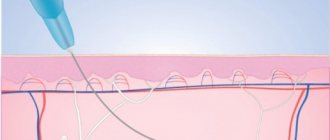
In most cases, injection chin surgery takes no more than 30 minutes. The cosmetologist treats the procedure area with anesthetic cream. Then he makes markings in accordance with the desired future shape and injects filler with a special syringe. Having introduced the filler, the doctor distributes it under the skin with massaging movements, creating the desired shape. The result will be noticeable within half an hour.
Chin correction and augmentation with fillers is virtually painless for most patients. But if you have increased sensitivity, instead of an anesthetic cream, the doctor may use lidocaine, novocaine or another anesthetic by injection.
PROCEDURE OF CHIN CONTOURING PROCEDURE
First of all, a detailed discussion of the expected parameters is carried out with a specialist. The cosmetologist then administers local anesthesia and performs a series of subdermal injections with an ultra-thin needle. Typically, chin correction with hyaluronic acid takes no more than an hour. According to reviews, chin contouring usually lasts for 6-18 months (drug manufacturers estimate 6-12 months), after which the procedure can be repeated. When using high-density fillers, the results last longer.
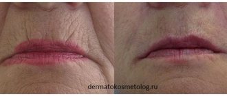
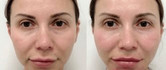
RESULTS: BEFORE AND AFTER PHOTOS
It is most convenient to evaluate the results of chin contouring using before and after photos. We remind you that photos are often taken before the procedure and immediately after - after 7-14 days the result may become even more pronounced and noticeable. A competent doctor follows the technique of performing the procedure and avoids dangerous areas, which provides the patient with the necessary level of safety. If necessary, after two weeks, additional correction of the selected zone can be carried out. Provided that the filler is chosen correctly, taking into account the characteristics of the skin, the patient’s age, and the desired results, an excellent effect from the procedure is guaranteed.
Results of the chin contouring procedure. Photos before and after
Egorova Margarita Leonidovna
Dermatovenerologist, cosmetologist, specialist in injection and thread techniques, Country Expert Teoxane Russia, Moscow
Carrying out basic aesthetic analysis, identifying morphological defects and involutional changes
Aesthetic medicine is called upon to solve two global problems in the facial area: harmonizing its proportions and improving the quality of the skin and subcutaneous tissues. Contour correction copes with the first task quite successfully. According to their functional purpose, all correction methods can be divided into adding, reducing and repositioning. Contour plastic surgery is classified as augmentative and is used to replenish the volume of the soft tissues of the face, although some modern drugs are at the intersection of correction and regeneration.
A frequent request from patients in the daily practice of a specialist is to eliminate jowls and improve the shape of the face in the chin area, angle and jaw line. The resources of contour correction to compensate for involutional changes in these areas are very limited and are applicable only in the initial stages of gravitational ptosis, with a slight displacement of the anterior and lower edges of the jowl to the point of forming furrows rather than folds, since in the latter case tissue reposition is required. Much more interesting and broader possibilities of the method can be used to compensate for morphological deficiencies in young patients and in the aspect of beauty in terms of leveling the surface of the face and restoring volume in its necessary areas.
Let's conduct a basic aesthetic analysis of the face and determine the goals of correction. This is quite easy to do without using the proportions of the golden section and the Marquardt and Fibonacci masks built on its basis, since any of us can easily distinguish harmonious from inharmonious, guided by unconscious perception.
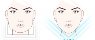
The height of the upper, middle and lower thirds of the face should be equal to each other ( Fig. 1 ). If the height in the lower third is less than necessary, it can be made larger by increasing the convexity of the chin into the lower plane of the face. The width of the face in the lower third is determined by a line drawn between the corners of the jaw. The proportional face has a narrowing in its upper third, widening in the middle and narrowing again in the lower, similar to the shape of a chicken egg turned upside down ( Fig. 2 ).
From the front view, it is worth determining the presence of zones of excessive depressions in the area of the labiomental, buccal-labial grooves and the anterior edge of the jowl.
The profile evaluates the length of the jaw line, the clarity of its angle, the degree of tissue displacement in the jowl area forward and down, the indentations in the area of its anterior and posterior edges, as well as the convexity of the chin in the anterior and lower plane of the face. The harmonious convexity of the chin in profile in its anterior plane should not protrude beyond the perpendicular drawn through the craniometric points of the nasospinal and pogonion ( Fig. 3 ).
Determining morphological and involutional defects also does not seem to be a difficult task, since any person who has nothing to do with aesthetic medicine can easily distinguish an old face from a young one and a harmonious one from an inharmonious one. A young and harmonious face has the correct shape, a uniform distribution of subcutaneous fatty tissue, and there are no areas of sharp depressions or skeletonization.
Signs of gravitational ptosis in the lower third of the face are visualized in the same way regardless of the morphotype of aging and are manifested by an increase and displacement of the convexity of the jowl with the compensatory formation of depressions in the adjacent areas ( Fig. 4 ).
Definition of correction tasks
When working with involutional changes, contour correction is effective only in the initial stage of gravitational ptosis. In such a situation, it is necessary to disguise the anterior (mandibular ligament) and posterior (platysma cutaneous ligament) edges of the jowl, visually lengthen the line of the lower jaw (distance from the craniometric points gonion and gnathion, Fig. 5 ), if necessary, enhance the clarity of its angle and increase the convexity of the chin. In addition, eliminate or reduce excess depressions in the area of the labiomental and buccal-labial grooves, mandibular ligament, platysma cutaneous ligament and ensure smooth interzonal transitions. Increasing the bulge in some areas of the face visually deepens others.
Young, harmonious faces have smooth interzonal transitions, so their restoration is always worth paying attention to when working with a patient of any age category. When harmonizing the parameters of the chin, pay attention to the deepening of the labiomental groove surrounding it from above, which is better visible in profile, and the zones of deepening of the mandibular ligament and buccal-labial groove located laterally from it ( Fig. 5 ) - with increasing convexity of the chin, they tend to visually deepen and most often also need to be filled out.
Even the most viscoelastic hyaluronic gel is not a solid substance and has significant plasticity (the ability to lose shape), so it is impossible to create a clear sculptural convexity in the shape of an angle from it, and it is not necessary, since a beautiful female face is not characterized by absolute geometric clarity and does not have sharp outlines in this zone. It is enough to distribute the gel linearly from the angle of the jaw along the line of its body, from the angle of the jaw along the line of its branch and fan-shaped between them. At the same time, the soft tissues in the area of the angle become more convex, and its clarity is more emphasized ( Fig. 4 ).
Anatomical structure and areas of potential danger
As in other areas of the face (with rare exceptions), the tissues in its lower third are formed by five layers: 1) skin, 2) superficial fat compartments, 3) superficial muscular aponeurotic system (SMAS), 4) deep cellular spaces (or their similarity) with the routes of vessels, nerves located in them, the beginning of the dense connective tissue of the ligamentous and septal apparatus of the face and 5) a layer that has variability and can be represented by the periosteum or deep fascia.
The anatomical substrate of the jowl at the superficial level is represented by the upper and lower jaw fat compartments. The superior one is located immediately below the nasolabial, lateral to the modiolus and borders the medial buccal. Due to its location below the nasolabial, it is visualized as its continuation. The lower jaw compartment is located below the upper jaw compartment, the mandibular ligament is located in its lower anterior corner, and laterally it borders on the temporobuccal fat compartment ( Fig. 6 ).
The appearance of the jowl is determined by the displacement of the skin and subcutaneous fat from the jaw line. But to a greater extent, its formation involves the deep anatomical space located under the SMAS in the lower part of the masseter, filled with branching fibers of the areolar tissue ( Fig. 7 ).
- The roof of the masseter space is the platysma (its lower plane), the bottom is the masseter fascia.
- The anterior border is represented by the masseter ligaments, the upper - by the septa, passing along a line drawn from the lower border of the tragus to the commissure of the lip.
- The posterior border is the place of transition of the platysma level (SMAS) into the platysmoauricular fascia (PAF), there are no spaces here, all tissues are tightly fused with each other and with the fascia of the anterior lobe of the parotid gland.
- The inferior border is a zone of weak attachment to the deep fascia of the masseter by membrane-like connective tissue.
The expansion and displacement of this space due to the weakness of the fixation of its roof (platysma) and the lower supporting membrane-like tissue is the cause of the appearance of jowls. In the anterior lower corner of the masseter space there is a fixation zone - the mandibular ligament; in the posterior lower corner the platysma cutaneous ligament begins. Between these connective tissue anatomical landmarks, the lower edge of the premasseteric space is poorly supported, causing it to move downward from the jaw line.
To a certain extent, the muscular mechanism also participates in the formation of jowls. With contraction of the muscles that depress the corners of the mouth, the anterior edges of the jowl are further displaced medially and protrude, resting against the mandibular ligament ( Fig. 8 ). When the platysma contracts in the area of its strings, the lower edge of the jowl additionally shifts downward between the mandibular ligament and the cutaneous ligament of the platysma. Taking these circumstances into account, better correction results are achieved in combination with the use of botulinum neuroprotein.
The mandibular facial nerve runs here in two branches. The superior one passes under the fascial capsule of the masseter and in front of the mandibular ligament rises outward to the subplatysmal level. The lower branch runs under the edge of the lower jaw ( Fig. 7 ).
The trigeminal nerve emerges from the mental foramen as part of the neurovascular bundle of the same name, located between the alveolar processes of the 1st and 2nd premolars. Along the line of the anterior edge of the masseter, the facial artery and vein enter the face through their incisura in the body of the lower jaw ( Fig. 9 ). The artery usually runs more medially, and in the middle third of the face its septum represents the morphological substrate of the nasolabial groove, and the vein runs more laterally and is responsible for the formation of the nasobuccal groove.
The right choice of drugs
Teoxane has been one of the top three manufacturers of hyaluronic fillers for 16 years and continues to invest its scientific research in developing products that provide ideal treatment results. The company's educational and methodological department is located in Geneva and collaborates with leading international aesthetic medicine specialists who generously share their knowledge and experience with colleagues. International experts at Teoxane recommend adhering to three basic principles in your work, which characterize the correct approach to working with a patient and are denoted by the abbreviation ATP (ANATOMY - TECHNOLOGY - PRODUCT), and always evaluate the patient’s face in statics and dynamics. Deep anatomical knowledge, informed selection of a product with optimal rheological properties and determination of the optimal technique are the key to a successfully performed procedure and a high degree of patient satisfaction with its result.
In independent studies, the Teosyal® UlrtaDeep PureSense static volumizer rightfully leads in its volumetric capacity and duration of action. This drug was created according to the classic reticulation patent, has high viscoelasticity and cohesiveness and can withstand compression deformation well in fixed areas of the face.
The disadvantage of static, highly reticular fillers is their insufficient ability to tolerate tensile deformation, against which they cease to be cohesive, begin to fragment and lose their shape. To preserve the naturalness of the face during facial expressions, Teoxane has developed the Teosyal® RHA line, the rheological properties of which are adapted to the natural biomechanics of facial tissues. Teosyal® RHA4 volumetric preparation retains elasticity (the ability to return to its original shape after deformation) when compressed and stretched and provides a natural appearance to the face during movement. Teosyal® RHA4 is created using the “preserved network” technology ( Fig. 10 ) - during its production, long chains of natural HA are preserved, capable of self-integrating into a three-dimensional mobile network. Inside the network, hyaluronic chains are interconnected by weak electrostatic bonds, which provide the property of maintaining shape when compressed and stretched. RHA's patented technology reduces the level of chemical modification, preserves the molecular weight of HA and creates gels that are elastic under tension and compression and are particularly well suited for injection into superficial, mobile fat compartments of the face to maintain its natural appearance during facial expressions ( Figure 11 ). The combination of layer-by-layer injection of static Teosyal® UlrtaDeep PureSense into deep fat compartments and dynamic Teosyal® RHA4 is used in the multi-layer (multi-layer) technique developed by Teoxane specialists to ensure ideal correction results.
Administration techniques
A young face is characterized by a smooth surface; a face with signs of aging shows an uneven relief of alternating zones of bulges and depressions. To replenish soft tissue volumes in young patients, it is usually sufficient to inject filler only into the deep compartments. In case of uneven facial relief in patients with involutional changes, injection only into deep compartments does not provide the maximum result of leveling its surface, and the procedure should be supplemented by injection of filler into superficial compartments using a multilayer technique ( Fig. 12 ).
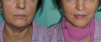
For clarity of understanding, the introduction techniques will be presented using the example of a 50-year-old patient, with a small degree of displacement of facial tissues in the jowl area forward and down, with a moderate degree of depression in the area of the mandibular ligament, in the areas of the buccal-labial and labiomental grooves. The angle of the jaw is quite well defined, due to its anatomically well-formed bony base and masseter tissue ( Fig. 13A ), and can only be slightly emphasized.
As a result of examination in profile, a slight deficiency in the convexity of the chin in the anterior plane of the face was revealed. To correct the convexity of the chin, the Teosyal® UlrtaDeep PureSense static volumizer was chosen.
The introduction was carried out through a 25G 40mm cannula. To enter the cannula along the lower edge of the chin, perpendicular to the skin, two entrance holes were made with a trocar to the depth of the subcutaneous fatty tissue. The cannula was located in deep fat (almost supraperiosteal). The filler was introduced into the premental space above the vestibular surface of the lower jaw, first slowly in a small portion to provide anesthesia, then until the cavity was completely filled.
Next, the depth of the labiomental groove was softened and the direction of insertion changed to the line of the lower jaw from the depression in the area of the mandibular ligament, which was carefully filled, to the beginning of the convexity of the chin. 1.2 ml of Teosyal® UlrtaDeep PureSense was used on each side in a ratio of 0.4 ml - 0.2 ml - 0.6 ml respectively.
To emphasize the angle of the jaw, Teosyal® RHA4 was used 1 ml on each side. The entrance hole was chosen on the anterior surface of the tissues, at the apex of the angle of the jaw. The introduction was carried out retrogradely at the level of superficial fat, first along the line of the jaw body from the posterior edge of the jowl to the angle of the jaw, then along the line of the jaw branch from the lower edge of the lobe to the angle of the jaw and then fan-shaped between these two lines ( Fig. 14 ). The area being corrected does not exhibit mobility, but Teosyal® RHA4 was chosen for angle correction due to its unique ability to remain cohesive (adhesion between drug particles) when filling large areas, tolerate any type of deformation and maintain ideal correction parameters for a long time.
In the static chin area and jawline from the mandibular ligament recess inclusive to the convexity of the chin, where injection was carried out under the SMAS, Teosyal® UlrtaDeep PureSense demonstrates its maximum volumetric capacity for a minimum of 1.5 years. As a result of the correction, the convexity of the chin was slightly increased, the identified depressions were filled and smooth interzonal transitions were restored (from the chin to the cheek, from the chin to the lip). The angle of the jaw became more pronounced and defined at the level of soft tissue ( Fig. 13B ).
Conclusion
The modern aesthetic medicine market provides specialists with all the conditions for successful and safe practice with high results from procedures. They have the opportunity to obtain fundamental knowledge of applied anatomy, various algorithms for safe and atraumatic drug administration based on aesthetic analysis of the face, and a wide selection of high-quality hyaluronic fillers that fully meet the requirements of the most demanding patients.
The article was published in the journal “Injection methods in cosmetology” No. 2-2020
As an advertisement
Recommended reading
Borzykh O.B. A combination of Teosyal UltraDeep and Teosyal RHA4 volumizers to create a line of ideal profile. Injection methods in cosmetology 2020; 1:26–31.
Egorova M.L. A dynamic method of creating volume in the zygomatic area using hyaluronic fillers Teosyal UltraDeep and Teosyal RHA4. The ideal combination to maintain a natural look to the face while it moves. Injection methods in cosmetology 2019; 4:32–38.
Mendelson B., Jacobson SR Surgical anatomy of the midcheek: facial layers, spaces, and the mid cheek segments. Clin Plast surg 2008; 35(3): 395–404.
Mendelson BC, Muzaffar AR, Adams WP Surgical anatomy of the midcheek and malar mounds. Plast Reconstr Surg 2002; 110(3): 885–896.
Rohrich RJ, Pessa JE, Ristow B. The youthful cheek and the deep medial fat compartment. Plast Reconstr Surg 2008; 121(6): 2107–2112.
CHIN CONTOUR: PRICE
The price for contour plastic surgery in St. Petersburg primarily depends on the type and volume of materials used, experience and skills of the specialist. The GMTClinic center offers fair prices for all types of contouring, while guaranteeing a high standard of service and absolute safety.
Let your face get a second wind of youth or create the oval face you have long dreamed of using chin contouring with fillers. Make an appointment with specialists by phone or using the online form.
If you want chin contouring, then at GMTClinic!