We continue to talk about rehabilitation after blepharoplasty. In the review article, we already touched on the topic of scars, but did not go into detail. Meanwhile, the timing of the disappearance of traces of the operation often worries patients the most.
Over the years of my practice, I have probably heard thousands of times the questions: “will the scar be very noticeable,” “how long will it take for the stitch to disappear,” “will the marks really remain for life,” and the like.
Today I will try to answer the most common ones and dispel your fears.
Scar healing time: misconceptions and facts
Many patients, knowing that the operation is easy, expect that the scars after blepharoplasty will disappear completely in 1-4 weeks. And if it doesn't happen as expected, some people start to panic. But plastic surgeons remind us that any scar or incision takes time to completely tighten and smooth out the scar on the surface of the skin.
And 4 weeks is too early to start worrying. After blepharoplasty, scars heal in several stages. It should take at least 10-12 weeks for them to completely smooth out and “even out” the color with the shade of the surrounding skin. This does not mean that the marks from the operation will be too noticeable for the entire 10 weeks. During this period, they can be masked using special cosmetics, and sometimes these actions are not necessary, since the scars quickly cease to be noticeable.
Lateral scars , located near the outer corner of the eye, are usually more noticeable than other blepharoplasty incisions. But their appearance also improves significantly after just a few weeks.
Thus, at least 2-3 months should pass .
Theory, diagnosis and clinical experience of correction.
Introduction
In terms of frequency, blepharoplasty ranks third in the world among all types of plastic surgery. It is second only to breast augmentation and liposuction. According to ISAPS (International Society of Aesthetic and Plastic Surgery), 1,099,960 blepharoplasty operations were performed worldwide in 2021 [11].
The most common indication for this intervention is age-related changes in the skin of the eyelids: gravitational ptosis (dermatochalasis), sagging of the skin of the upper eyelids and the formation of a double contour of the lower eyelid, downward displacement of the level of the eyebrow line.
This surgical procedure has a number of contraindications: increased arterial or intraocular pressure, dry eyes, chronic diseases of the thyroid gland and cardiovascular system in the stage of decompensation, diabetes mellitus, cancer, disorders of the blood coagulation system.
The aesthetic indicators of aging of the periorbital area are directly dependent on the condition of the eyelids and surrounding tissues. In this area, the skin primarily undergoes age-related changes, loses tone and sags. Deformations of the eyelids can cause severe functional disorders, and they also represent a clear cosmetic defect. These conditions can be the consequences of congenital pathologies, inflammatory diseases, injuries and burns, senile changes, and postoperative complications.
Overhanging eyelid skin in older people often leads to a narrowing of the visual field. Eyelid surgery can expand the area of peripheral vision or partially reduce intraocular pressure. Drooping upper eyelids may be due to excess skin, hypertrophy of the orbicularis oculi muscle, herniated fatty tissue, or a combination of these.
The rate of healing of a postoperative wound after blepharoplasty depends not only on the skill of the surgeon, the extent of the surgical intervention, but also on the patient himself. If the integrity of the skin is damaged, the healing process immediately starts, which ends with tissue restoration.
The classic version of acute wound healing is a complex, dynamic and perfectly planned process, consisting of four main sequential and at the same time overlapping stages, regulated by several factors: hemostasis, inflammation, proliferation, remodeling [12].
The last stage of the healing process begins with the development of granulation tissue and is the longest in time. As the matrix matures, the amount of fibronectin and hyaluronan decreases, and the collagen fiber bundles increase in diameter, which helps to increase the tensile strength of the wound. However, newly formed collagen fibers achieve only 80% of the strength of intact skin.
Remodeling is a delicate balance between tissue formation and degradation, controlled by the activity of proteolytic enzymes, mainly matrix metalloproteinases (MMPs) and their natural tissue inhibitors [4]. Disruption or slowdown of this process, dysfunction of the extracellular matrix can lead to the formation of pathological scars.
In molecular biology, the extracellular matrix (ECM) is defined as a complex network formed by numerous structural macromolecules (proteoglycans, collagens, elastin). By interacting with each other and with cells, these structural macromolecules maintain the structural integrity of tissues [9].
It is the matrix that provides an organized environment within which migrating cells can move and interact with each other. All macromolecules that make up the ECM are produced by cells in the matrix. The most important component of the extracellular matrix is a gel-like environment formed by proteoglycans - extremely elongated polypeptide chains with numerous polysaccharide chains of glycosaminoglycans attached through covalent bonds.
Numerous chains of proteoglycans are attached to a special type of glycosaminoglycan, a polymer of hyaluronic acid called hyaluronan. Its threads hold the gel structure together into a single whole, and this polysaccharide “gel” can resist the compression and stretching of the ECM and at the same time provide rapid diffusion of nutrients, building materials and hormones between the blood and connective tissue cells, providing regulatory and plastic functions, especially relevant for postoperative wound healing.
Mechanically, the gel structure is reinforced using different types of fibers:
- fibers that form the skeleton of connective tissue,
- flexible fibers that give connective tissue elasticity,
- mesh fibers that form cross-links between all other fibers and connect all other components of the fabric.
To date, 29 types of collagen have been described, the function of which is not fully established. There are 9 types of collagen fibers in the skin [5], which give connective tissue strength and durability. Each collagen fiber is several micrometers in diameter and consists of thousands of individual collagen polypeptide chains tightly packed together.
Collagens are among the most abundant proteins in the extracellular matrix and connective tissue. In the human genome, there are about 50 genes encoding various collagens, and the products of these genes form about 30 types of collagen fibers found in a wide variety of tissues. Excess collagen fibers or too little collagenase activity lead to increased fiber density and the formation of less flexible tissue. On the contrary, excessive collagenase activity will lead to uncontrolled fragmentation of collagen, which will make the tissue more amorphous. Any disruption of this complex multicomponent process entails a failure in the healing of the wound and can cause the formation of pathological scars.
Currently, there is no clear distinction between the terms “scar” and “scar tissue.” The term “scar,” used in a number of sources, is a connective tissue formation that is formed during the healing process of a wound, and scar tissue appears in its last phase—the epithelialization phase [1, 8].
On the other hand, all components of the dermis are connective tissue formations, and all repair processes accompanied by scar formation occur within the connective tissue of the dermis, while defects within the epidermis are not accompanied by scarring.
It is more correct to talk not about “replacement with connective tissue,” as is done in some manuals, but about disruption of the processes of intercellular interaction during connective tissue repair, leading to the formation of pathological scars.
The reliable reasons for the development of hypertrophic and keloid scars have not yet been established. However, it is known that the formation of pathological scars is based on disturbances in the synthesis of structural components of the dermis, with intensification of the formation of collagen fibers, an increased influence of growth factors on angiogenesis, fibroblast proliferation, increased activity of metalloproteinase inhibitors and changes in normal fibroblast apoptosis [8]. In case of dysregulation of the processes of synthesis, assembly, breakdown of collagen, elastin and glycosaminoglycans (GAGs), insufficient or, conversely, excessive scarring may be observed.
Before surgical treatment, it is advisable to evaluate the reparative potential of the skin, as well as the genetically determined mechanisms responsible for subsequent tissue remodeling. Currently, molecular genetic methods are being actively introduced into the practice of cosmetologists and plastic surgeons, making it possible to analyze probable risks. After conducting a DNA test, for example, “Cosmetology” from BGG [Fig. 1], before the procedures, you can build a program that takes into account the individual risk of formation of defective collagen, reduced or increased activity of matrix metalloproteinases and will allow you to avoid contraindicated procedures and / or additionally prepare before blepharoplasty.
It is advisable to include in complex therapy drugs that increase the regenerative potential of the dermis (PRP, placenta preparations, collagen, GAGs and others), normalizing collagenogenesis and the synthesis of other components of the dermal matrix.
Authors: Natalya Bychkova, Ph.D., cosmetologist of the highest category, Izhevsk. Irina Bykova, cosmetologist, Izhevsk.
You can read the article in Oblik magazine. Esthetic guide No. 2(35), pp. 46-47
How do scars heal after blepharoplasty?
In the first 4 weeks after blepharoplasty, scars, although it is more accurate to call them postoperative scars, go through a granulation phase, when new, “young” connective tissue, rich in small vessels, forms at the site of the incision. As a result, by the end of the first month, the incision site turns into a pink scar. Over the next month and a half, the scar is organized, turning it into a thin white line that barely protrudes above the surface of the skin.
In rare cases where scars are healing too slowly, your doctor may recommend revision of the surgical scar to eliminate excess connective tissue growth.
Principle of procedure
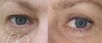
A laser machine is a station that produces light. A laser beam is a beam of light consisting of strictly ordered waves clearly directed parallel to each other. It is this orderliness that allows the resulting beam to be used in medicine and cosmetology. Each of the chromophores (water, melanin, hemoglobin) is capable of absorbing laser radiation of its own wavelength and becoming a target during cosmetic laser treatment of the skin. Adjustable wavelength and frequency allow the cosmetologist to selectively influence the structure of the scar without harming other components of the epidermis. Laser eyelid resurfacing: before and after photos
Why so expensive?
Laser resurfacing, the price of which can reach several tens of thousands for a set of procedures, is an expensive cosmetology service. The fact is that the production and purchase of a laser system costs a lot of money. Accordingly, procedures performed on expensive equipment cannot be cheap. In the production of laser systems for both cosmetology and “big” medicine, artificially grown precious stones and complex multi-level systems of optical mirrors are used. The laser installation consists of three blocks:
- The source of energy is the supply of electricity.
- The working fluid is an element for converting energy (electricity) into a beam of light of a certain type of wave.
- An optical system of mirrors is necessary for repeated reflection of radiation; it sets the frequency and wavelength of the output beam.
Optical mirror systems in laser systems allow you to set the wavelength and frequency of radiation necessary to correct a specific skin defect. If the wave frequency is not calibrated correctly, you can get serious complications or, conversely, not get the desired effect.
The name of the laser depends on the working fluid used:
| Laser name | Working fluid | Output wavelength, nanometers | Chromophore effects and scope of application |
| Carbon dioxide (CO2) | Mixture of gases | The laser beam at the output is in the infrared range, wavelength up to 10600 nm | Affects mainly water. Widely used for laser resurfacing of scars, including after blepharoplasty. |
| Erbium (Erbium YAG) | Erbium in yttrium aluminum garnet (gemstone) | 2940 nm | Impact chromophore - water, used for laser resurfacing of scars |
| Neodymium (Nd:YAG) | Yttrium aluminum garnet (gemstone) | 1064 nm | Affects hemoglobin and melanin, not used for resurfacing eyelids after blepharoplasty |
| KTP laser | Potassium Titanyl Phosphate | 532 nm | Affects hemoglobin and melanin, not used for resurfacing |
| Alexandrite | Alexandrite crystals (gemstone) | 755 nm | The gold standard is laser hair removal, but not resurfacing. |
The cost of one scar resurfacing procedure depends on its size. On average, laser resurfacing of a small scar after blepharoplasty will cost from 600 to 1,500 rubles. If the operation was performed on the lower and upper eyelids, laser resurfacing will cost from 2,400 to 6,000 rubles per session.
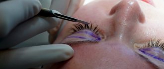
Caring for seams
The doctor makes the incisions necessary for the operation directly on the eyelids. The sutures run close to the eyeball. Therefore, if an infection enters the wound, the entire eye may be damaged. Complications can affect the condition of individual parts of the eye and vision.
Proper care of sutures will help minimize the risk of complications.
In the first days after surgery, you must wear a sterile bandage. It will protect the seam from possible infection and particles of dust and dirt.
In addition, you need to treat the seams with an antiseptic and special ointments. The doctor will prescribe the necessary medications for you based on the medical history collected before the procedure.
After the stitches are removed, your specialist may recommend that you use a gel that helps restore the damaged area.
Release forms
Pharmacy chains sell Dermatix and Dermatix Ultra gels. The cost of the drugs is almost the same, while the “Ultra” version contains vitamin C. At the same time, there is a version of silicone dressings, popularly called “Dermatix patch”. Attempts to purchase Dermatix ointments or creams are doomed to failure; such forms of release do not exist.
The drug is sold in pharmacy chains without prescriptions, although its availability is questionable. The cost of gels is surprising with a big difference, it is rational to compare the options before purchasing.
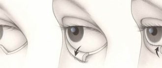
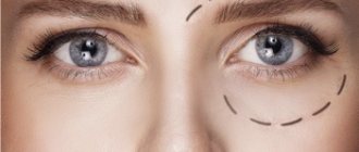
Who is suitable for circumferential blepharoplasty?
Circular blepharoplasty can eliminate congenital or acquired pathologies of the periorbital eye area in one go. Indications for it are:
- Sagging and drooping corners of the eyes due to aging
- Ptosis of soft tissues in the area of the upper and lower eyelids simultaneously
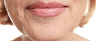
- Deep expression lines and folds around the eyes
- Acquired defects
- Bags and swelling under the eyes along with sagging of the upper eyelid
- Fat hernias
- Another indication may be a congenital predisposition to sagging eyelid skin at an early age or dissatisfaction with appearance without medical indications.
Immediately after surgery
The doctor applies a bandage to the stitches. The eyelids remain swollen for the first few days . There may be slight bruising on the surrounding skin. The sutures are removed (more precisely, the threads are removed from the seams) after 3-7 days. If the surgeon worked with absorbable threads, then this is not required; such material disappears on its own within the same period. During the first week, the areas where the incisions were made remain very sensitive and should not be touched without the permission of a healthcare professional. All necessary procedures (treatment and dressing) will be performed by a doctor or nurse.
Your task is not to touch, pull or rub your eyelids..